SCALEVIEW-A2
Dr. Atsushi Miyawaki et al. developed a water-based optical clearing reagent, Scale, which clears fixed biological samples withoutquenchingfluorescent protein. Using SCALEVIEW-A2, bodily tissues can be cleared of their opacity to provide a clearly defined image. The SCALEVIEW approach integrates seamlessly with multiphoton microscopes, providing the power to visualise 3-dimensional structures at unprecedented depths in morphologically intact tissue.
Optical characteristic : Refractive index ne(20℃) of SCALEVIEW-A2 is 1.377 - 1.381.

Protocol
Example: Rendering mouse brain tissue transparent for deep imaging according to reference1).
- Transcardially perfuse an anesthetised mouse with 4% paraformaldehyde (PFA)/PBS (pH 7.5~8.0).
- Remove the whole brain and subject it to post-fixation in 4% PFA/PBS at 4 °C for 10 hrs and cryo-protection in 20% sucrose/PBS at 4°C for 24 hrs.
- Embed the sample in OCT compound and freeze it with liquid nitrogen.
- Thaw and rinse the sample in PBS, and fix it again with 4% PFA/PBS for 20 min at room temperature.
- Clear the sample by incubation in SCALEVIEW-A2 at room temperature.
More than 30 ml of SCALEVIEW-A2 is required for an adult mouse brain.
A full week of incubation may be necessary for transparency.
During incubation, stir slowly using an orbital shaker.
Exchanging SCALEVIEW-A2 each day accelerates the transparency process.
The sample might be expanded 10-30% in one direction after incubation - Perform deep imaging of the transparent brain using an appropriate dipping objective lens.Use SCALEVIEW-A2 as the immersion medium.
- The transparent brain can be stored for a long time in SCALEVIEW-A2 at 4°C.
1.Image of a mouse brain after SCALEVIEW-A2 process
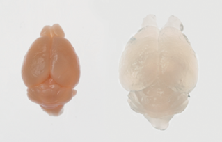
Figure 1
Amouse brain after SCALEVIEW-A2 process
Left: a mouse brain before SCALEVIEW-A2 process
Right: a mouse brain after SCALEVIEW-A2 process.
2. Imaging data using SCALEVIEW-A2
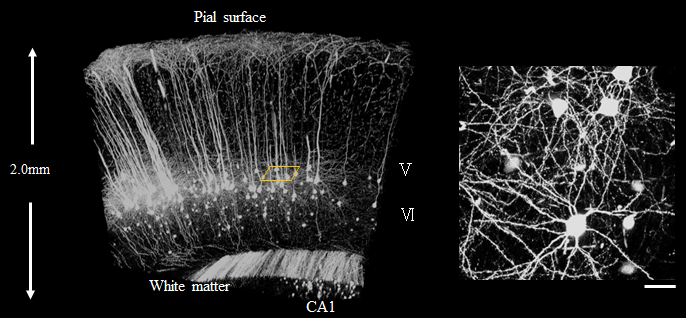
Figure 2
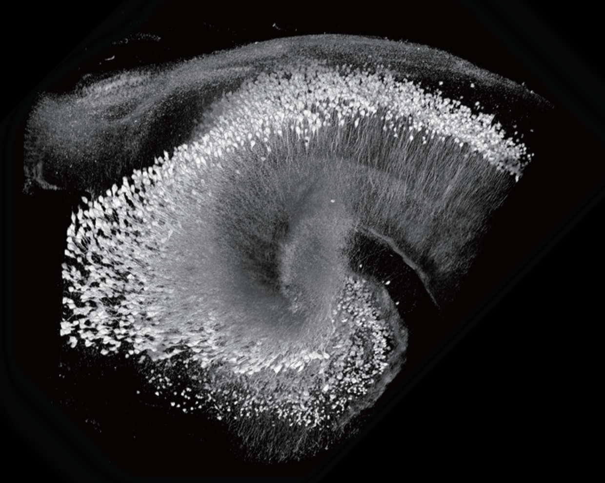
Imaging of Thy1-YFP(H Line) mouse brain after SCALEVIEW-A2 process using Multiphoton microscope and Multiphoton dedicated objective :OLYMPUS, model:XLPLN10XSVMP.Bars, 50μm.
Image
Imaging data using SCALEVIEW-A2
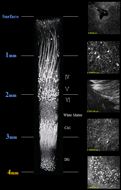
Figure 3
Deep imaging of Thy1-YFP(H Line) mouse brain after SCALEVIEW-A2 process using Multiphoton microscope and Multiphoton dedicated objective :OLYMPUS,
model:XLPLN25XSVMP(NA1.0 WD4mm).
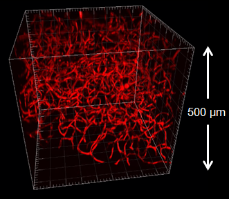
Figure 4
Image of mouse pancreas(labeling process the blood vessel using Lectin Texas Red) after SCALEVIEW-A2 process using Confocal microscope and objective :OLYMPUS, UPLSAPO30XS..
Reference
1) Hama,H.et al. : Nature Neuroscience 14, 1481(2011).
Product List
- Open All
- Close All
For research use or further manufacturing use only. Not for use in diagnostic procedures.
Product content may differ from the actual image due to minor specification changes etc.
If the revision of product standards and packaging standards has been made, there is a case where the actual product specifications and images are different.