Exosome Flow Cytometry Kits


It is said that exosomes are difficult to be analyzed by flow cytometry, due to their very small size (30 - 100 nm). Fujifilm Wako developed a flow cytometry kit for exosome analysis by combining Tim4 protein, which specifically binds to phosphatidylserine on the surfaces of extracellular vesicles, including exosomes, and magnetic beads. This kit enables easy and sensitive flow cytometry analysis of exosomes without purification.
Product Line-up
More Information
Can Exosomes Be Analyzed Using Flow Cytometry?
Flow cytometry (FCM) is a technique that aligns cells in a narrow flow path, sequentially irradiates them with a laser, and detects the resulting scattered light and fluorescence. This method is widely used in the analysis of heterogeneous cell populations as it allows for rapid measurement of the characteristics of individual cells.
Extracellular vesicles (EVs), including exosomes, are known to be heterogeneous, and flow cytometry analysis is expected to be a valuable method for analyzing the characteristics and composition of exosomes. However, the diameter of exosomes is around 100 nm, which presents a challenge for analysis under conditions typically designed for cell sizes on the μm scale. On the other hand, there have been reports of successful flow cytometry analysis of exosomes by optimizing various factors, including such parameters as detection limits, the concentration of exosomes, controls for comparison, and staining methods for labeling exosomes.
Although flow cytometry of exosomes is feasible, the diverse conditions employed by researchers have been problematic in terms of experiment reproducibility and interpretation of results. To address this issue, Joshua et al. published a position paper in 2020, on flow cytometry analysis of EVs, titled “MIFlowCyt-EV1).” This paper outlines a framework for optimizing various conditions, including pre-processing of samples, setting control parameters, and calibration of the flow cytometer. Consequently, researchers considering flow cytometry analysis of EVs are advised to refer to this paper.
Application of PS Affinity Method in Flow Cytometry
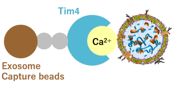
Fujifilm Wako, in collaboration with Professor Hanayama of the Department of Immunology, Kanazawa University Graduate School of Medical Sciences, has developed a novel exosome isolation and purification method called the “PS affinity method,” utilizing Tim4, a protein that specifically binds to phosphatidylserine (PS) on the exosome surface2). This method captures exosomes through PS-Tim4 binding and is theoretically applicable to flow cytometry. However, the challenge was that the diameter of the magnetic beads used in the isolation and purification kit was too small, thus falling below the detection limit of flow cytometry.
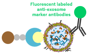
To address this issue, Fujifilm Wako modified the diameter of the magnetic beads and the composition of the washing buffer, optimizing them for flow cytometry analysis. This led to the successful development and commercialization of the “PS Capture™ Exosome Flow Cytometry Kit” (Product Number: 297-79701). This kit isolates exosomes using magnetic beads and labels them with the desired fluorescent labeled anti-exosome marker antibodies (e.g., anti-CD9, anti-CD63, anti-CD81 antibodies) (Figure 1). By flowing the exosomes while they remain bound to the magnetic beads, it is now possible to detect exosomal surface antigens using flow cytometry*.
*Note that in this kit, multiple Tim4 molecules are bound to each bead particle. As the exact number of Tim4 molecules per bead particle cannot be guaranteed, quantitative analysis using this kit is not recommended.
1) Isolation of exosomes
Exosomes are isolated from the sample
by Exosome Capture Beads.
2) Staining of exosomes
Exosomes are stained with fluorescent-labeled
anti-exosome marker antibodies.
3) Flow cytometry analysis
Flow cytometry analysis is performed
with exosomes bound to magnetic beads.
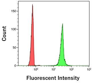
Figure 1 Assay principle of PS Capture™ Exosome Flow Cytometry Kit
References
- Welsh, J. A. et al.: J. Extracell. Vesicles, 9(1), 1713526(2020).
MIFlowCyt‐EV: A framework for standardized reporting of extracellular vesicle flow cytometry experiments - Nakai, W. et al.: Sci. Rep., 6(1), 33935(2016).
A novel affinity-based method for the isolation of highly purified extracellular vesicles
For research use or further manufacturing use only. Not for use in diagnostic procedures.
Product content may differ from the actual image due to minor specification changes etc.
If the revision of product standards and packaging standards has been made, there is a case where the actual product specifications and images are different.