Antibodies (Cancer)
As cancer treatments shift from chemotherapy to personalized therapy using molecular targeted cancer drugs, the scope of molecular biological research on cancer is accelerating. Antibody experiments have contributed greatly to elucidate physiological phenomena in cancer cells by molecular biological methods. Fujifilm Wako offers high-performance antibodies against molecules, including asialo GM1, integrin, IDH, and podoplanin, that are important in cancer research.
More Information

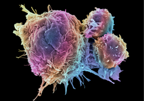
Cancer cells (malignant tumor, malignant neoplasm) grow out of normal control, invade (infiltrate) surrounding tissues, and metastasize to other organs and tissues.
Different from normal cells, cancer cells have the following characteristics:
- Can proliferate indefinitely
- Proliferate, survive, and divide independently of signals from surrounding cells
- Are less likely to commit suicide by apoptosis
- Have a high gene mutation rate
- Are highly invasive
- Can proliferate and survive in organs and tissues to which they have metastasized
Normal cells transform into cancer cells when gene mutations have accumulated and these properties are acquired. Genes that cause cancer when over-activated are called oncogenes, and genes that cause cancer when inactivated are called tumor-suppressor genes.
Pathologic Classification of Gliomas and Factors Involved
Gliomas are cancers that arise from glial cells (neuroglia) and are the most frequent (~ 30%) among primary brain tumors. 1)
In the WHO Classification of Tumours of the Central Nervous System, revised 4th edition published in May 2016, molecular diagnosis based on the molecular classification of tumor tissues is nearly mandatory for pathologic classification of gliomas. Gliomas are classified based on the presence or absence of mutations in IDH (isocitrate dehydrogenase) 1, IDH2, ATRX, or p53 gene, as well as chromosome 1p/19q codeletion (loss of the short arm of chromosome 1 and the long arm of chromosome 19).
Mutation in the promoter region of TERT (telomerase reverse transcriptase) gene is reported as a mutation found in almost all gliomas with IDH gene mutation and 1p/19q codeletion.
Cancer and Fibrosis
Fibrosis is a phenomenon in which the connective tissue proliferates abnormally. The basis of fibrosis is an excessive accumulation of extracellular matrix (ECM), such as collagen produced by fibroblasts. As wound tissues and tumors share a common fibrosis formation process, tumors have long been called "non-healing wounds."2)
In wound tissues, lymphocytes are activated by inflammatory response. The activated lymphocytes release growth factors and cytokines that promote fibril formation to activate macrophages and fibroblasts. Fibroblasts differentiate into myofibroblasts and produce ECM such as collagen. Normally, the wound is healed during this process, but excessive production of ECM by myofibroblasts due to chronic injury or inflammation leads to fibrosis.3) TGF-β plays a major role in fibrosis. Integrin is involved in the activation of TGF-β,4) and CTGF is involved in the differentiation to myofibroblasts and the promotion of collagen synthesis by TGF-β.5)
In tumors, fibrosis is promoted by an increase of myofibroblasts, which produce ECM, and remodeling enzymes involved in ECM reorganization, cross-linking, and hardening.6)
Cancer and Platelet Aggregation
Platelets are a blood cell component that play an important role in hemostasis through blood coagulation. Based on previous research, it is known that platelet aggregation is accelerated when cancer cells enter blood vessels from primary lesions and metastasize to other tissues through the blood stream (hematogenous metastasis).
The aggregated platelets cover cancer cells, making them less susceptible to attack by the immune system,7) and preventing their physical destruction while flowing in blood vessels. Also, it has been clarified that platelets promote adhesion of cancer cells to the blood vessels to which they have metastasized and that growth factors (such as VEGF) released by activation of platelets cause an increase in vascular permeability and angiogenesis necessary for invasion and proliferation of cancer cells8) to greatly contribute to metastasis of cancer cells.
Podoplanin is one of factors involved in platelet aggregation by cancer cells. It is a type I transmembrane protein and is extensively glycosylated, and causes platelet aggregation by binding to the podoplanin receptor CLEC-2 on platelets.9) As its expression is increased in many cancer cells, podoplanin has been used as a tumor marker. Anti podoplanin antibodies with neutralizing activity are expected as a therapeutic drug to suppress cancer metastasis and deterioration due to cancer.
References
- National Cancer Center Japan:
https://www.ncc.go.jp/jp/ri/division/brain_tumor_translational_research/project/020/20170907102931.html (Apr. 2, 2021) - Dvorak, H. F.: Cancer Immuno. Res., 3(1), 1(2015).
Tumors: wounds that do not heal-redux - Wynn, T. A.:J. Pathol., 214(2), 199(2008).
Cellular and molecular mechanisms of fibrosis - Biernacka, A., Dobaczewski, M., and Frangogiannis, N. G.:Growth Factors, 29(5), 196(2011).
TGF-β signaling in fibrosis - Duncan, M. R., et al.: FASEB J., 13(13), 1774(1999).
Connective tissue growth factor mediates transforming growth factor beta-induced collagen synthesis: down-regulation by cAMP - Piersma, B., Hayward, M. K. and Valerie M. W.:Biochim. Biophys. Acta. Rev. Cancer, 1873(2), (2020).
Fibrosis and cancer: A strained relationship - Placke, T. et al.: Cancer Res., 72(2), 440(2012).
Platelet-derived MHC class I confers a pseudonormal phenotype to cancer cells that subverts the antitumor reactivity of natural killer immune cells - Goel, H. L. and Mercurio, A. M.: Nat. Rev. Cancer, 13(12), 871(2013).
VEGF targets the tumour cell - Suzuki-Inoue, K. et al. : J. Biol. Chem., 282(36), 25993(2007).
Involvement of the snake toxin receptor CLEC-2, in podoplanin-mediated platelet activation, by cancer cells
For research use or further manufacturing use only. Not for use in diagnostic procedures.
Product content may differ from the actual image due to minor specification changes etc.
If the revision of product standards and packaging standards has been made, there is a case where the actual product specifications and images are different.


