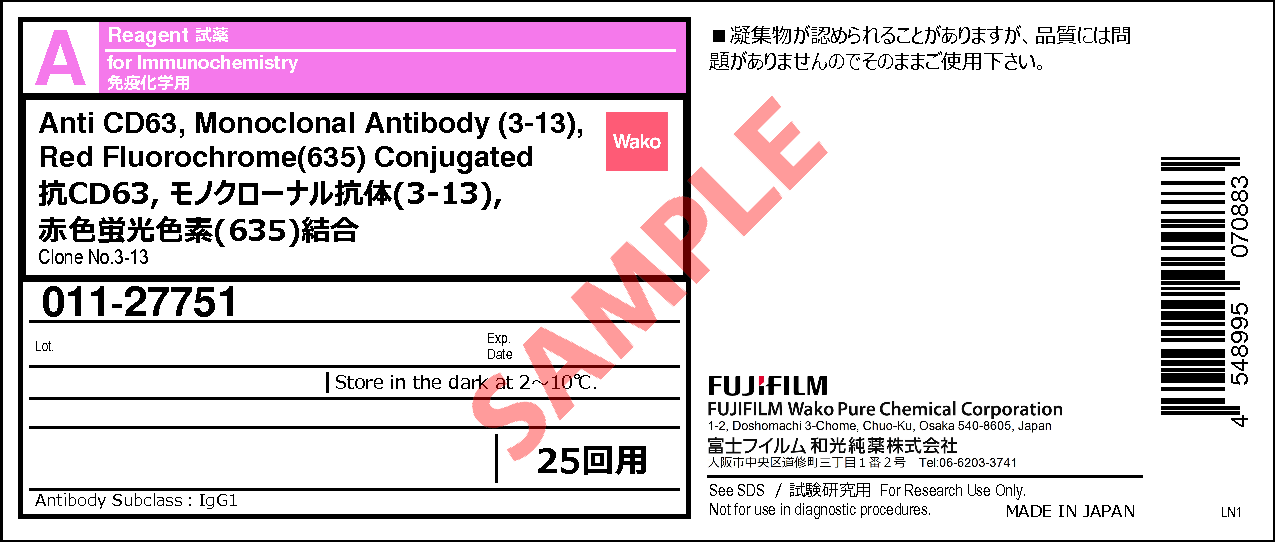
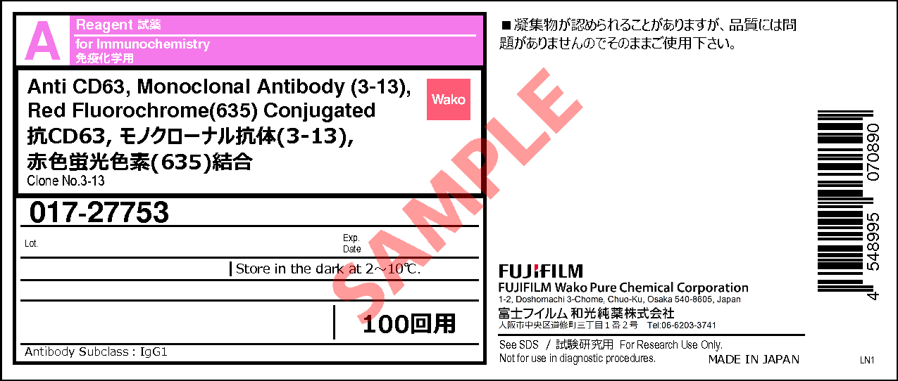
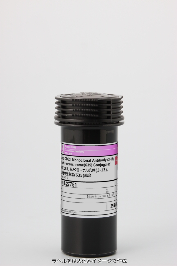
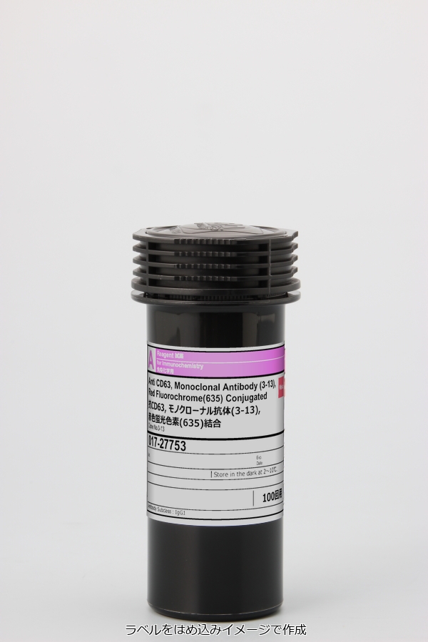
Anti CD63, Monoclonal Antibody (3-13), Red Fluorochrome(635) Conjugated
- for Immunochemistry
- Manufacturer :
- FUJIFILM Wako Pure Chemical Corporation
- Storage Condition :
- Keep at 2-10 degrees C.
- Structural Formula
- Label
- Packing
- SDS
|
Comparison
|
Product Number
|
Package Size
|
Price
|
Availability
|
Certificate of Analysis
|
Purchase |
|---|---|---|---|---|---|---|
|
|
|
25Tests
|
|
Out of Stock |
||
|
|
|
100Tests
|
|
Out of Stock |
※Check availability in the US with the distributor.
Document
Product Information
| Cross Reactivity | Human [Note] This antibody doesn’t recognize the mouse, rat and bovine CD63. |
|---|---|
| Clone No. | 3-13 |
| Host | Mouse |
| Subclass | IgG1 |
| Formulation | 1×TBS aqueous solution with Tween 20, the stabilizer, and preservative |
| Label | Red Fluorochrome(635) Excitation: 634 nm, Emission: 654 nm |
▼Evaluating a reactivity
The reactivity of this antibody against a serum of each organisms was measured by ELISA.
When the signal value was the same level as the blank, it was judged as "non-reactive".
Application
Flow Cytometric analysis of exosomes: 10uL (1 test) product for 33uL or more culture supernatant/biofluids os recommended for the exosome flow cytometry using PS Capture Exosome Flow Cytometry Kit [code No. 297-79701].
Please optimize the most appropriate concentration for your analysis and samples.
Application Data
Flow cytometry
Ex / Em = 634 nm / 654 nm
FCM analysis of COLO201 cells
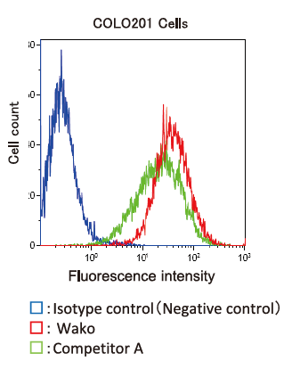
Flow cytometric analysis of COLO201 cells was performed using Wako (3-13) and competitor A.
Sample amount: 1 x 10⁶ cells
Antibody amount: 10 μL (1 test)
Detection of CD63 on the surface of exosome in COLO201 cell culture supernatant and human serum
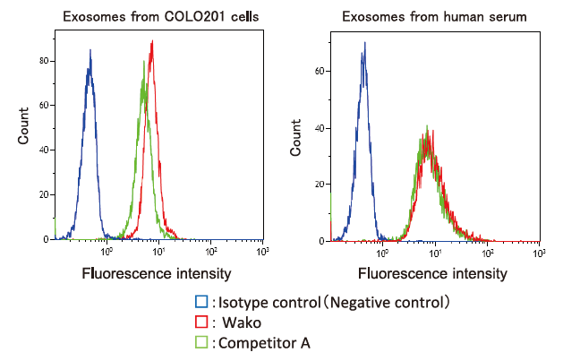
From pretreated※ COLO201 cell conditioned medium (8-fold dilution) and human serum, exosomes were isolated using PS Capture™ Exosome Flow Cytometry Kit (Code No. 297-79701) and analyzed by flow cytometry using this antibody or the other company product.
※ Pretreatment condition: 10,000 x g, 30min.
Sample amount (supernatant / serum): 33 μL
This product achieved detection of CD63 on the COLO201 cell surface and exosome surface comparable to or more sensitive than the other company product.
Overview / Applications
| Outline | Anti human CD63 (lysosome-associated membrane glycoprotein 3 : LAMP3) mouse monoclonal antibody was produced by hybridoma, clone number; 3-13. CD63 is the four-transmembrane protein and used for the marker protein for activated platelet. Recently, CD63 is also used for a marker protein for extracellular vesicles, which contain proteins, DNAs, mRNAs, microRNAs and are expected as a novel biomarker. This product is antibody conjugated red fluorochrome (635) which is similar to Cy5 and FUJIFILM Wako's original compound to Anti CD63, Monoclonal Antibody (3-13). It is suitable for Flow Cytometry experiment. Excitation : 634 nm, Emission : 654 nm |
|---|---|
| Purpose | Flow Cytometry |
| Instructions | Flow Cytometric analysis of exosomes: 10uL (1 test) product for 33uL or more culture supernatant/biofluids os recommended for the exosome flow cytometry using PS Capture Exosome Flow Cytometry Kit [code No. 297-79701]. Note: The particle size of exosome is too small to detect by Flow Cytometry. To using Anti CD63, Monoclonal Antibody(3-13), Red Fluorochrome conjugated, please use with the exosome capture beads. Please optimize the most appropriate concentration for your analysis and samples. Flow cytometric analysis of cells: 10uL (1 test) product for 1 x 10^6 cells. |
Property
| Specificity | Specific for human CD63. *This antibody doesn't recognize the mouse, rat, and bovine CD63. [Reactivity confirmation condition] The reactivity of these antibodies against the serum of each organisms was measured by ELISA. When the signal value was the same level as the blank, it was judged as "non-reactive. |
|---|---|
| Composition | 1 x TBS aqueous solution with Tween 20, the stabilizer, and the preservative. |
Manufacturer Information
Alias
For research use or further manufacturing use only. Not for use in diagnostic procedures.
Product content may differ from the actual image due to minor specification changes etc.
If the revision of product standards and packaging standards has been made, there is a case where the actual product specifications and images are different.


