


Intestinal Organoids
Intestinal organoids are three-dimensional tissues that recapitulate intestinal tissue. Rather than consisting of a single cell type induced from ES or iPS cells, they are formed from multiple cell types. Because they allow researchers to observe human intestinal development in vitro, they are increasingly used in disease-mechanism studies, such colorectal cancer research, and in drug safety testing.
Learn about the basics of intestinal organoids, the principles behind their generation, directed differentiation protocols, and the reagents used.

Intestinal Organoids
The intestine is a complex organ that, beyond absorbing nutrients, plays key roles in immune function and in maintaining homeostasis. Yet human intestinal development and disease mechanisms are difficult to study directly, and research to date has relied mainly on animal models and cultured cells.
Human intestinal organoids have emerged in recent years as a way to address these challenges. Generated from human iPS or ES cells, these three-dimensional tissues recapitulate the structure, function, and development of the human intestine in vitro. As such, they hold promise as a next-generation model for intestinal research.
Intestinal organoids are formed by differentiating human pluripotent stem cells into definitive endoderm and then modulating signals such as WNT and FGF in a stepwise manner. The cells are first directed toward midgut-like tissue, forming human intestinal organoids (HIOs). Activating BMP signaling then confers colonic characteristics, forming human colonic organoids (HCOs). When the resulting spheroids are cultured in a matrix for about one month, HIOs and HCOs develop a tissue structure in which the epithelium is surrounded by mesenchymal cells. The epithelium contains enterocytes, paneth cells, goblet cells, enteroendocrine cells, and other cell types. This differentiation and maturation process recapitulates intestinal formation during embryogenesis, making these organoids an excellent model for understanding human intestinal development.
Whereas conventional adult tissue-derived organoids consist mainly of epithelial cells, iPS cell-derived intestinal organoids contain both epithelial and mesenchymal cells. This allows cell–cell interactions and tissue formation processes to be examined under conditions that more closely reflect physiological reality. These organoids also avoid the species differences that limit animal models, allowing researchers to study human-specific physiology and gene expression.
Intestinal organoids are now widely used in studies of intestinal development, congenital gastrointestinal disorders, and diseases such as colorectal cancer. In addition, patient-derived iPS cells and patient-derived organoids (PDOs) are increasingly being used in drug screening and personalized medicine. More recently, co-culture with immune cells and enteric neurons, along with organ-on-a-chip technology, is being used to build intestinal models that more closely mimic the native intestine.
Intestinal organoids thus serve as an innovative research platform, supporting a wide range of applications, from understanding human intestinal development, physiology, and disease mechanisms to drug discovery and regenerative medicine. Further advances are expected in the years ahead.
Methods for Generating Intestinal Organoids
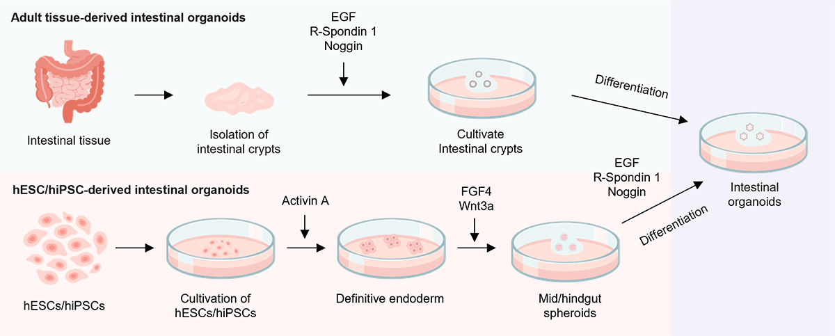
There are two major approaches to generating intestinal organoids. The first method involves isolating crypts from patient-derived intestinal tissue and culturing them to form organoids.
The second method uses human pluripotent stem cells, such as human ES cells (hESCs) or human iPS cells (hiPSCs), which are guided through stepwise differentiation to produce intestinal organoids.
Protocol for the Generation of Intestinal Organoids
We present protocols and recommended reagents for generating intestinal organoids.
Please refer to the references for detailed protocol information. (Tominaga, K. et al., 2024 , Liu, F. et al., 2025)
Protocol examples 1 (Tominaga, K. et al., 2024)
| iPSCs | Definitive endoderm | Mid/hindgut Endoderm | Mid/hindgut spheroids | |||
|---|---|---|---|---|---|---|
 |
||||||
| Media | hPSC medium | RPMI | Advanced DMEM/F12 | |||
| Serum/Additives | NEAA/FBS | N2/B27/L-Glutamine/HEPES/PS | ||||
| Cell Dissociation Reagents | Accutase | |||||
| Scaffolding Materials | EHS gel matrix | EHS gel matrix | ||||
| Small Molecules | Y-27632 | Y-27632 | ||||
| CHIR99021 | ||||||
| Cytokine | ActivinA | FGF4 | EGF | |||
| Equipment | 6-well plate | 24-well plate | 24-well plate | |||
*Created based on data from Tominaga, K. et al., 2024
Protocol examples 2 (Liu, F. et al., 2025)
| iPSCs | Endoderm-mesoderm co-induction | Intestinal patterning | Intestinal maturation | |||
|---|---|---|---|---|---|---|
 |
||||||
| Media | hPSC medium | RPMI/hPSC medium | RPMI | Advanced DMEM/F12 | ||
| Serum/Additives | L-Ala-L-Gln/B27-insulin | L-Ala-L-Gln/B27/N2 | ||||
| Cell Dissociation Reagents | Trypsin-EDTA | |||||
| Scaffolding Materials | EHS gel matrix | |||||
| Small Molecules | A83-01/5AZA/PD98059/DAPT | |||||
| Cytokine | BMP4/ActivinA | Wnt3a/FGF4 | EGF/R-Spondin 3/Noggin | EGF | ||
| Equipment | 96-well plate | |||||
*Created based on data from Liu, F. et al., 2025
References
For research use or further manufacturing use only. Not for use in diagnostic procedures.
Product content may differ from the actual image due to minor specification changes etc.
If the revision of product standards and packaging standards has been made, there is a case where the actual product specifications and images are different.