


Cardiac Organoids
Cardiac organoids are three-dimensional tissues that partially recapitulate cardiac tissue. Rather than consisting of a single cell type induced from ES or iPS cells, they are formed from multiple cell types. Because they allow researchers to observe the processes of human heart development in vitro, they are being used in disease research, including studies of human fetal cardiogenesis and disease-specific signaling abnormalities.
Learn about the basics of cardiac organoids, the principles behind their generation, directed differentiation protocols, and the reagents used in their preparation.
Cardiac Organoids
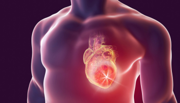
Cardiovascular disorders, including cardiovascular disease (CVD) and congenital heart defects (CHD), are the leading cause of death worldwide, accounting for an estimated 17.9 million deaths annually. Despite this, the development of organoids for cardiovascular disease research has lagged behind that for other organs such as the kidney, brain, and intestine, possibly due to the structural complexity of the heart.
To date, various cardiac models, including animal models and spheroid cultures, have been used to study disease mechanisms and support drug discovery. However, many of these models consist of only specific cell types and cannot fully recapitulate the multicellular interactions—including those involving epicardial and endocardial cells—or the structural complexity of the organ in vivo.
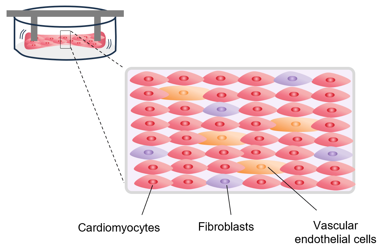
To address these challenges, researchers developed 3D cardiac tissue (engineered heart tissue), which is created from a mixture of individually differentiated cardiomyocytes, vascular endothelial cells, fibroblasts, and other cells. 3D cardiac tissue excels at modeling the mechanical and electrophysiological properties of tissue, and numerous disease models have been reported for recapitulating the phenotypes of various heart diseases and for drug discovery research. However, because this approach is based on the artificial assembly of differentiated cells, it cannot readily reproduce the natural processes of early embryonic heart development or the accompanying formation of the endogenous extracellular matrix (ECM).
In contrast, cardiac organoids, a field that has attracted growing interest in recent years, form through the self-organization of stem cells during cardiac differentiation. As a result, their structure more closely resembles that of the actual heart, including naturally formed layers. With current technology, 3D cardiac tissue is built from cells that are already relatively well differentiated, whereas the cells in cardiac organoids often remain at early stages of development. Because cardiac organoids more closely recapitulate in vivo developmental processes, they can be used to model malformations by introducing specific genetic mutations or altering culture conditions. They are therefore expected to become a more effective platform for elucidating the mechanisms of heart development and studying developmental disorders—areas that have been difficult to investigate using conventional models.
Cardiac Organoids vs. 3D Cardiac Tissue
| Cardiac Organoids | 3D Cardiac Tissue | |
|---|---|---|
| Structure | 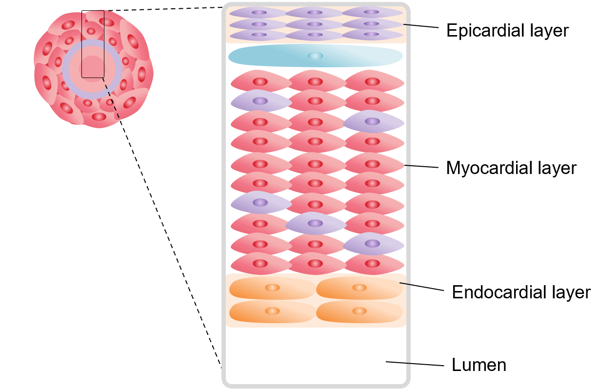
|
|
| Generation | Formed through spontaneous self-organization | Formed by combining differentiated cells on scaffolds or in molds |
| Disease Modeling | Mimic developmental processes, making them well suited for modeling embryonic development and developmental abnormalities | Models of various hereditary and non-hereditary diseases have been reported |
| Variability | High tissue-to-tissue variability | Low tissue-to-tissue variability |
Protocol for the Generation of Cardiac Organoids
We present protocols and recommended reagents for generating cardiac organoids.
Please refer to the references for detailed protocol information. (Tian, Y. et al., 2022 , Voges, HK. et al., 2023 )
Protocol examples 1 (Tian, Y et al., 2022)
| iPSCs | Differentiation of cardiac organoids | ||||||||
|---|---|---|---|---|---|---|---|---|---|
 |
|||||||||
| Media | hPSC Medium |
RPMI | |||||||
| Serum/Additives | B27-insulin | B27-insulin | |||||||
| Cell Dissociation Reagents | Accumax | ||||||||
| Small Molecules | Y-27632 | CHIR99021 | Wnt-C59 | CHIR99021 | |||||
| Cytokine | BMP4/ ActivinA |
||||||||
| Equipment | 96-well plate |
||||||||
*Created based on data from Tian, Y. et al., 2022
Protocol examples 2 (Voges, HK. et al., 2023)
| iPSCs | Differentiation of cardiomyocytes and stromal cells |
Cell dissociation | Cardiac organoid culture | ||||||||
|---|---|---|---|---|---|---|---|---|---|---|---|
 |
|||||||||||
| Media | hPSC medium | RPMI | MEMα | MEMα | DMEM-glucose | ||||||
| Serum/Additives | B27-insulin/PS/L-Ascorbic Acid/L-Ala-L-Gln | B27/PS/L-Ascorbic Acid /L-Ala-L-Gln | PS/L-Ascorbic Acid/FBS/L-Ala-L-Gln | PS/L-Ascorbic Acid/B27/ L-Ala-L-Gln | PS/L-Ascorbic Acid/B27-insulin/ L-Ala-L-Gln/Aprotinin/D-glucose/Palmitic Acid | ||||||
| Insulin | |||||||||||
| Cell Dissociation Reagents | Collagenase I/Trypsin-EDTA | ||||||||||
| Scaffolding Materials | EHS gel matrix | Collagen I /EHS gel matrix/10×DMEM |
PDMS | ||||||||
| Small Molecules | CHIR99021 | IWP-4 | |||||||||
| Cytokine | BMP4/ActivinA/FGF2 | PDGF-BB/FGF2 | |||||||||
| Equipment | T25 flask | T25 flask | 96-well plate | ||||||||
*Created based on data from Voges, HK. et al., 2023
References
For research use or further manufacturing use only. Not for use in diagnostic procedures.
Product content may differ from the actual image due to minor specification changes etc.
If the revision of product standards and packaging standards has been made, there is a case where the actual product specifications and images are different.