Iba1 항체
| 고객님의 편의를 위해 이 페이지는 기계 번역으로 제공됩니다. 정확한 번역을 제공하기 위해 최선을 다하고 있으나, 기계 번역은 완벽하지 않을 수 있습니다. 기계 번역된 내용이 만족스럽지 않으신 경우, 영어 버전 을 확인해 주시기 바랍니다. |
Iba1(이온화 칼슘 결합 어댑터 분자 1)은 중추 신경계의 미세아교세포에서만 특이적으로 발현되므로 미세아교세포 표지자로 사용됩니다. 말초 조직에서는 대식세포에서 발현되며 AIF-1로도 알려져 있습니다. 후지필름 와코의 "Anti Iba1, Rabbit (면역세포화학용)"(제품 번호 019-19741)은 전 세계 연구자들이 미세아교세포 마커 항체의 표준으로 사용하고 있습니다. 또한, 후지필름 와코는 "Anti Iba1, Rabbit Monoclonal Antibody (6A4), recombinant"(제품 번호 018-28523)을 개발했는데, 이 제품은 Anti Iba1, Rabbit (for immunocytochemistry)과 동일한 성능을 가지고 있습니다.
Iba1 소개
Iba1(이온화 칼슘 결합 어댑터 분자 1)은 약 17 kDa의 칼슘 결합 단백질입니다. 이 단백질은 중추 신경계의 미세아교세포에서 특이적으로 발현되기 때문에 미세아교세포 마커로 사용됩니다1). 이 단백질은 휴지 상태와 활성화된 미세아교세포 모두에서 발현되지만, 활성화된 미세아교세포에서 더 높은 발현 수준을 보이는 것으로 보고되었습니다2). 또한 말초 조직의 대식세포에서도 발현되며, AIF-1(이종 이식 염증 인자-1)로 알려져 있다.
Iba1은 세포 내 F-액틴에 결합하여 액틴 다발을 형성한다. 액틴 다발의 형성은 세포 이동 및 식작용 과정에서 관찰되는 세포막 주름 현상에 필수적인 것으로 여겨진다3).
후지필름 와코의 Anti-Iba1 항체
후지필름 와코의 “Anti Iba1, 토끼 (면역세포화학용)” (제품 번호 019-19741)는 면역조직화학 염색을 통해 미세아교세포의 돌기까지 염색할 수 있어, 전 세계 연구자들이 미세아교세포 표지 항체의 표준으로 사용하고 있습니다.
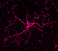
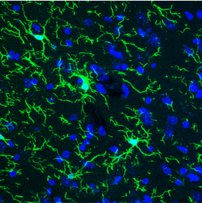
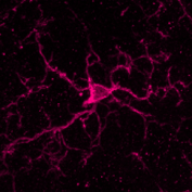
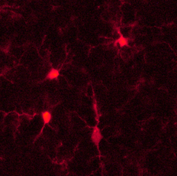
마우스 소뇌의
면역조직화학 염색
Anti Iba1, Rabbit
(for immunocytochemistry)
간 출판물 수
토끼 유래 Anti-Iba1 항체(면역세포화학용)를 다룬 논문이 점점 더 많이 발표되고 있으며, 이 중에는 주요 학술지(Nature, Cell, Neuron 등)에 게재된 논문도 다수 포함되어 있고, 연간 관련 논문 수는 1,500편을 넘어섰다.
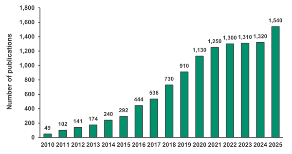
Google Scholar에서 “Iba1 019-19741 Wako”라는 키워드로 검색했습니다.
| 저널 | 발표 논문 수 | 2024년 영향력 지수 |
|---|---|---|
| Nature | 49 | 50.5 |
| Cell | 43 | 45.5 |
| Science | 3 | 44.7 |
| Nature Medicine | 19 | 58.7 |
| Nature Neuroscience | 89 | 21.2 |
| Nature Immunology | 13 | 27.7 |
| Nature Biotechnology | 9 | 33.1 |
| Nature Methods | 4 | 36.1 |
| Nature Biomedical Engineering | 6 | 26.8 |
| Nature Cell Biology | 3 | 17.3 |
| Nature Genetics | 1 | 31.7 |
| Neuron | 67 | 14.7 |
| Total | 306 |
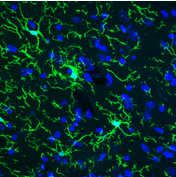
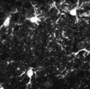
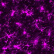
마우스 대뇌 피질의
면역조직화학 염색
Anti Iba1, Rabbit
Monoclonal Antibody (6A4), recombinant
토끼 단일클론 항체 개발
2023년, 후지필름 와코는 토끼 단일클론 항체인 “Anti Iba1, 토끼 단일클론 항체 (6A4), 재조합”을 출시했습니다. (제품 번호 018-28523)를 출시했습니다. 당사는 이 항체가 쥐와 랫트 뇌의 면역조직화학 염색에서 “Anti Iba1, 토끼 (면역세포화학용)”과 동일한 성능을 보인다는 사실을 확인했습니다. 또한 쥐 망막의 면역조직화학 염색에서도 우수한 결과를 보인 것으로 보고되었습니다(응용 데이터 참조).
제품 라인업
선정 흐름도

*2 Abbreviations:IHC(F)=Immunohistochemistry (frozen section), IHC(P)=Immunohistochemistry (paraffin section), ICC=Immunocytochemistry; WB=Western blotting
Product Lineup
| 진행자 | Rabbit | Goat | Mouse | ||||||||
|---|---|---|---|---|---|---|---|---|---|---|---|
| 제품 번호 | 018-28523 | 019-19741 | 013-27691 | 016-20001 | 016-26461 | 015-28011 | 012-28401 | 013-26471 | 011-27991 | 016-26721 | 013-27593 |
| 설명 | Rabbit monoclonal antibody | For Immno- staining (Standard) | For paraffin section | For western blotting | Biotin-conjugated | SPICA Dye™ 568-conjugated | SPICA Dye™ 594-conjugated | Red fluorochrome (635)-conjugated | Goat polyclonal antibody | Mouse monoclonal antibody (NCNP24) | Mouse monoclonal antibody (NCNP27) |
| 접사 | Unconjugated | Unconjugated | Unconjugated | Unconjugated | Biotin | SPICA Dye™ 568 Ex=556 nm Em=591 nm |
SPICA Dye™ 594 Ex=575 nm Em=611 nm |
Red fluorochrome (635) Ex=634 nm Em=654 nm |
Unconjugated | Unconjugated | Unconjugated |
| 항체 | Monoclonal antibody | Polyclonal antibody | Monoclonal antibody | ||||||||
| Concentration (mg/mL) | 1.0-1.2 | 0.5-0.7 | 0.5-0.7 | 0.5-0.7 | 0.5-0.6 | 0.5-0.6 | 0.5-0.6 | 0.5-0.6 | 0.6-0.7 | 0.9-1.6 | 0.9-1.3 |
| 항원 | Synthetic peptide (Iba1 C-terminal sequence) | ||||||||||
| Cross-reactivity | Mouse Rat |
Mouse Rat Other species also reported*1 |
Mouse Rat |
Human Mouse Rat |
Marmoset Mouse Rat |
Mouse Rat |
Mouse Rat |
Mouse Rat |
Mouse Rat |
Marmoset Mouse Rat |
Human |
| 신청*2 수치는 권장 농도를 나타냅니다 | IHC(F) 1:200-10,000 FCM 1:100-10,000 |
IHC(F) 1:500-1,000 ICC 1:500-1,000 |
IHC(P) 1:500-1,000 |
WB 1:500-1,000 |
IHC(F) 1:200-2,000 |
IHC(F) 1:200-2,000 |
IHC(F) 1:200-2,000 |
IHC(F) 1:200-2,000 |
IHC(F) 1:250-1,000 IHC(P) 1:250-1,000 WB 1:1,000 |
IHC(F, DAB) 1:500-2,000 IHC(F, fluorescent) 1:100 |
IHC(P, DAB) 1:100-1,000 |
| 권 | 100 μL | 50 μg | 50 μg | 50 μg | 100 μL | 100 μL | 100 μL | 100 μL | 100 μL | 50 μL | 50 μL |
*2 Abbreviations:IHC(F)=Immunohistochemistry (frozen section), IHC(P)=Immunohistochemistry (paraffin section), ICC=Immunocytochemistry; WB=Western blotting
Iba1 항체를 이용한 미세아교세포 면역조직화학 검사의 표준 프로토콜
후지필름 와코의 “Anti Iba1, 토끼 (면역세포화학용)” (제품 번호 019-19741)은 미세아교세포의 돌기 부분까지 염색할 수 있어 우수한 미세아교세포 표지 항체입니다. 여기서는 미세아교세포 면역조직화학 검사를 수행할 때의 프로토콜과 주의사항을 설명합니다. 예시로 마우스 뇌의 냉동 절편과 형광 염색제를 사용합니다.
1.조직 절편 제작
1-1. 생쥐에 관류 처리를 한 뒤 4% 파라포름알데히드-인산염 완충액으로 고정한다.
1-2. 자당으로 대체하고, 냉동 블록을 준비한다.
1-3. 마이크로톰을 사용하여 두께 50 µm의 조직 절편을 준비한다.
2.세탁 - 블록킹
2-1. 0.3% 트리톤 X-100/PBS 용액으로 5분씩 3회 세척합니다.
2-2. 1% BSA, 0.3% Triton X-100이 포함된 PBS 용액에서 실온에서 2시간 동안 차단 처리한다.
다음의 블로킹 용액도 사용할 수 있습니다:
· 1% BSA, 0.3% Tween-20/PBS
· 2차 항체 숙주의 정상 혈청 3%
3.1차 항체 반응
3-1. 1% BSA, 0.3% Triton X-100/PBS 용액에 Anti-Iba1, 토끼 항체(면역조직화학용)를 1:1,000 희석하여 첨가합니다.
3-2. 4°C에서 하룻밤 동안 배양한다.
4.세탁
4-1. 0.3% 트리톤 X-100/PBS 용액으로 5분씩 3회 세척합니다.
5.2차 항체 반응
5-1. 형광 표지된 항토끼 IgG 항체(예: Jackson ImmunoResearch, 제품 번호 111-545-144)를 1% BSA, 0.3% Triton X-100/PBS 용액에 1:1,000 희석하여 첨가합니다.
5-2. 실온에서 2시간 동안 배양합니다.
6.세탁
6-1. 0.3% 트리톤 X-100/PBS 용액으로 5분씩 3회 세척합니다.
7.장착
7-1. 부품을 고정 매체에 고정하십시오.
8.관찰
8-1. 형광 현미경이나 공초점 현미경으로 해당 단면을 관찰하십시오.
왼쪽에 설명된 기법으로 미세아교세포가 잘 염색되지 않는 경우, 조직 절편 제작 후 다음 방법 중 하나를 사용하여 항원 회수 처리를 수행하십시오:
(A) 시트르산 완충액(pH 6.0)을 사용하여 90°C에서 9분간 처리
(B) TE 완충액(pH 9.0)을 사용하여 90°C에서 9분간 처리
응용 프로그램 데이터
면역조직화학
▼Anti Iba1, Rabbit Monoclonal Antibody (6A4), recombinant
Species: Mouse
Site: Cerebral cortex
Sample: Frozen section
Antibody concentration: 1:1,000
Species: Mouse
Site: Retina
Sample: Frozen section
Antibody concentration: 1:2,000
Data by courtesy of
Dr. Watanabe, Dr. Iwagawa
University of Tokyo Hospital
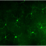
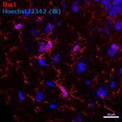
▼Anti Iba1, Rabbit (for immunocytochemistry)
Species: Rat
Site: Cerebral cortex
Sample: Frozen section
Antibody concentration: 1:1,000
Data by courtesy of
Dr. Sanagi, Dr. Manabe,
Dr. Ichinohe, Dr. Kohsaka
National Center of
Neurology and Psychiatry
Species: Mouse
Site: Cerebellum
Sample: Frozen section
Antibody concentration: 1:1,000

- Species
- Mouse
- Site
- Cerebral cortex/Area postrema (Medulla oblongata)
- Sample
- Frozen section
- Antibody concentration
- P2RY12 → Anti P2RY12, Guinea Pig (Product No. 011-28873) 1:900
Iba1 → Anti Iba1, Rabbit (for Immunocytochemistry) 1:500
Laminin → Anti Laminin Antibody (Rat, Made in Dr. Miyata’s Lab) 1:200
Data by courtesy of
Dr. Miyata, Department of Applied Biology, Kyoto Institute of Technology
[결과]
Iba1 양성 세포 중 뇌 실질에 존재하는 세포들은 P2RY12 양성이며, 소교세포로 추정된다(화살표 머리). 반면, 혈관 기저막 주변에는 Iba1 양성/P2RY12 음성 세포가 존재하며, 이 세포들은 대식세포로 추정된다(화살표).
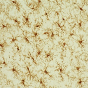
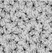
▼Anti Iba1, Rabbit (for paraffin section)
Species: Rat
Site: Hippocampus vicinity
Sample: Paraffin section
Antibody concentration: 1:1,000
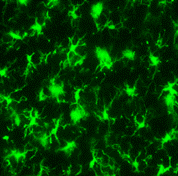
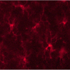
▼Anti Iba1, Rabbit, Biotin-conjugated
Species: Rat
Site: Cerebral cortex
Sample: Frozen section
Antibody concentration: 1:200
Data by courtesy of
Dr. Sanagi, Dr. Manabe,
Dr. Ichinohe, Dr. Kohsaka
National Center of
Neurology and Psychiatry
▼Anti Iba1, Rabbit, SPICA Dye™ 568-conjugated
Species: Rat
Site: Cerebral cortex
Sample: Frozen section
Antibody concentration: 1:200
▼Anti Iba1, Rabbit, SPICA Dye™ 594-conjugated
Species: Rat
Site: Cerebral cortex
Sample: Frozen section
Antibody concentration: 1:200
▼Anti Iba1, Rabbit, Red Fluorochrome (635)-conjugated
Species: Rat
Site: Cerebral cortex
Sample: Frozen section
Antibody concentration: 1:200
Data by courtesy of
Dr. Sanagi, Dr. Manabe,
Dr. Ichinohe, Dr. Kohsaka
National Center of
Neurology and Psychiatry
Species: Mouse
Site: Hippocampus
Antibody concentration: 1:200
Data by courtesy of
Dr. Takata
Division of Integrated
Pharmaceutical Sciences,
Kyoto Pharmaceutical University
▼Anti Iba1, Mouse Monoclonal Antibody (NCNP24)
Species: Rat
Site: Cerebral cortex
Sample: Frozen section
Antibody concentration: 1:1,000
Data by courtesy of
Dr. Sanagi, Dr. Manabe,
Dr. Ichinohe, Dr. Kohsaka
National Center of
Neurology and Psychiatry
Western blotting
▼Anti Iba1, Rabbit (for Western Blotting)
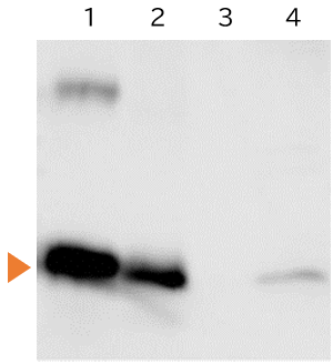
1.Iba1 protein...10 ng
2.Rat Microglia...10 μg
3.Rat Neuron...10 μg
4.Rat cerebral cortex...150 μg
SDS-PAGE: 5.5% stacking gel, 12.5% running gel, 100V
Blocking: 3% skim milk/TBS, 1 hour, room temperature.
Primary antibody: 1:1,000 concentration, 3% skim milk/TTBS, overnight, 4℃
Secondary antibody: Peroxidase-labeled Anti Rabbit IgG (1/5,000), 3% skim milk/TTBS, 1 hour, room temperature
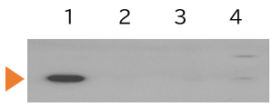
1.Rat primary culture microglia...10 μg
2.Rat primary culture neuron...10 μg
3.Rat primary culture astrocyte...10 μg
4.Rate cerebral cortex...100 μg
Primary antibody: Anti Iba1, Goat, 1:1,000
Secondary antibody: Anti Goat IgG, HRP-conjugated
참고 문헌
- Imai, Y., Ibata, I., Ito, D., Ohsawa, K. & Kohsaka, S.: Biochemical and biophysical research communications, 224(3), 855(1996).
A Novel Geneiba1 in the Major Histocompatibility Complex Class III Region Encoding an EF Hand Protein Expressed in a Monocytic Lineage - Mori, I., Imai, Y., Kohsaka, S. & Kimura, Y.: Microbiology and immunology, 44(8), 729(2000).
Upregulated expression of Iba1 molecules in the central nervous system of mice in response to neurovirulent influenza A virus infection - Sasaki, Y., Ohsawa, K., Kanazawa, H., Kohsaka, S. & Imai, Y.: Biochemical and biophysical research communications, 286(2), 292(2001).
Iba1 is an actin-cross-linking protein in macrophages/microglia. - Zhao, S. et al.: Cell, 180(4), 796(2020).
Cellular and Molecular Probing of Intact Human Organs - Ahn, J.H. et al.: Lab. Anim. Res., 28(3), 165 (2012).
Comparison of alpha-synuclein immunoreactivity in the spinal cord between the adult and aged beagle dog - Ide, T. et al.: J. Vet. Med .Sci., 72(1), 99 (2010).
Histiocytic Sarcoma in the Brain of a Cat - Gaige, S. et al.: Neurotoxicology, 34, 135(2013).
c-Fos immunoreactivity in the pig brain following deoxynivalenol intoxication: Focus on NUCB2/nesfatin-1 expressing neurons - Rodriguez-Callejas, J.D. et al.: Front. Aging Neurosci., 8, 315(2016).
Evidence of Tau Hyperphosphorylation and Dystrophic Microglia in the Common Marmoset - Fantin, A. et al.: Blood, 116(5), 829(2010).
Tissue macrophages act as cellular chaperones for vascular anastomosis downstream of VEGF-mediated endothelial tip cell induction
Product List
- Open All
- Close All
Anti-Iba1, 토끼 (단일클론 항체)
Anti-Iba1, 염소 (다클론 항체)
Anti-Iba1, 마우스 (단일클론 항체)
Anti-Iba1, 마우스 (단클론 항체)
For research use or further manufacturing use only. Not for use in diagnostic procedures.
Product content may differ from the actual image due to minor specification changes etc.
If the revision of product standards and packaging standards has been made, there is a case where the actual product specifications and images are different.
The prices are list prices in Japan.Please contact your local distributor for your retail price in your region.