Anti Iba1, Rabbit Monoclonal Antibody (6A4), recombinant
- for Immunochemistry
- Manufacturer :
- FUJIFILM Wako Pure Chemical Corporation
- Storage Condition :
- Keep at -20 degrees C.
- GHS :
-

-
 Close
Close
Close
Close -
 Close
Close
- Structural Formula
- Label
- Packing
- SDS
|
Comparison
|
Product Number
|
Package Size
|
Price
|
Availability
|
Certificate of Analysis
|
Purchase |
|---|---|---|---|---|---|---|
|
|
|
20uL
|
|
In stock in Japan |
||
|
|
|
100uL
|
|
In stock in Japan |
※Check availability in the US with the distributor.
Document
Product Overview
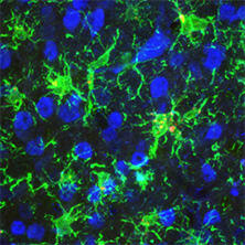
Iba1 (Ionized calcium-binding adapter molecule 1) is an approximately 17 kDa calcium-binding protein. It is used as a microglial marker because it is expressed specifically in microglia in the central nervous system1). It is expressed in both resting and activated microglia, but is reportedly expressed more highly in activated microglia2). It is also expressed in macrophages in peripheral tissues and is known as AIF-1 (Allograft inflammatory factor-1). Iba1 binds to F-actin in cells to form actin bundles. The formation of actin bundles is thought to be required for the membrane ruffling observed during cell migration and phagocytosis3).
Anti Iba1, Rabbit Monoclonal Antibody (6A4), recombinant is a rabbit monoclonal antibody that has the same performance as "Anti Iba1, Rabbit (for immunocytochemistry)" (Product Number 019-19741) in immunohistochemical staining of mouse and rat brains. It has also been reported to obtain good results in immunohistochemical staining of mouse retinas.
Features
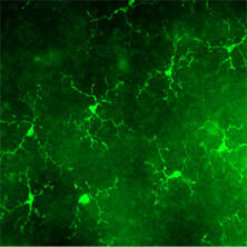
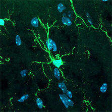
- Enables even the processes of microglia to be immunohistochemical stained.
- Same performance as "Anti Iba1, Rabbit (for immunocytochemistry)".
- Relatively small amount needed, with concentrations ranging from 1:200 to 1:10000.
- Can be used for immunohistochemical staining of mouse retinas.
Antibody Information
| Clonality | Monoclonal |
|---|---|
| Antigen | Synthetic peptide (Iba1 C-terminal sequence) |
| Host | Rabbit |
| Buffer | PBS (50% Glycerol), 0.05% NaN3 |
| Concentration (mg/mL) | 1.0 - 1.2 |
| Conjugate | Unconjugated |
| Cross-reactivity | Mouse, Rat |
| Application | Immunohistochemistry (Frozen Section) 1:200 - 10,000 Flow cytometry 1:100-10,000 |
Data
Performance Data
Comparison with our conventional product (rabbit polyclonal antibody)
Polyclonal Antibody
(Product No. 019-19741)
Monoclonal Antibody
(Product No. 018-28523)
Species: Rat
Site: Cerebral cortex
Sample: Frozen section
Antibody concentration: 1:200
Data by courtesy of
Dr. Nakajima, Faculty of Science and Engineering, Soka University
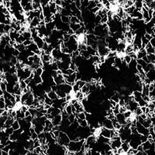
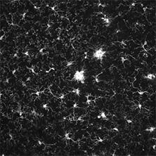
Comparison with company A (rabbit monoclonal antibody)
Company A
(rabbit, monoclonal)
Fujifilm Wako
(Product No. 018-28523)

Species: Mouse
Site: Cerebral cortex
Sample: Frozen section
Antibody concentration: 1:1,000
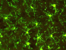
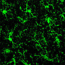
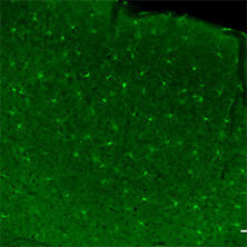
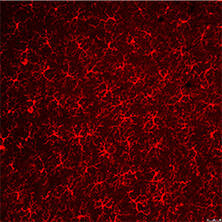
Application Data
Immunohistochemistry
Mouse
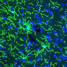
Species: Mouse
Site: Cerebral cortex
Sample: Frozen section
Antibody concentration: 1:1,000
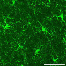
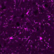
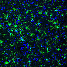
Species: Mouse
Site: Cerebral cortex
Sample: Frozen section
Antibody concentration: 1:200
Data by courtesy of
Dr. Koizumi, Interdisciplinary Graduate School of Medicine,
University of Yamanashi
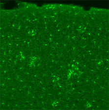
Species: Mouse
Site: Cerebral cortex
Sample: Frozen section
Antibody concentration: 1:200
Note: Preparation of frozen sections → PFA fixation
Data by courtesy of
Dr. Ino, Graduate School of Medicine, Osaka University
Species: Mouse
Site: Cerebral cortex
Sample: Frozen section
Antibody concentration: 1:1,000
Data by courtesy of
Dr. Nonaka, Graduate School of Engineering, Kyoto University
Species: Mouse
Site: Cerebral cortex
Sample: Frozen section
Antibody concentration: 1:1,000
Data by courtesy of
Dr. Ito, Graduate School of Medicine, Juntendo University
Species: Mouse
Site: Cerebral cortex
Sample: Frozen section
Antibody concentration: 1:1,000
Data by courtesy of
Dr. Tsuruta and Mr. Ueda, Faculty of Life and Environmental Sciences, University of Tsukuba
Species: Mouse
Site: Cerebral cortex
Sample: Frozen section
Antibody concentration: 1:1,000
Data by courtesy of
Dr. O, S University
Species: Mouse
Site: Cerebral cortex
Sample: Frozen section
Antibody concentration: 1:1,000
Data by courtesy of
Dr. Y, Institute of R
Species: Mouse (APPNL-F/NL-F)
Site: Cerebrum
Sample: Fixed tissue slice
Antibody concentration: 1:200
Note: Tissue clearing with Scale method
Data by courtesy of
Dr. Hama, RIKEN Center for Brain Science
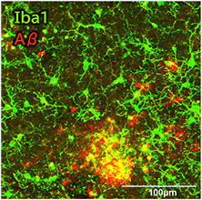
Species: Mouse (Model of Alzheimer Disease)
Site: Cerebral cortex
Sample: Frozen section
Antibody concentration: 1:500
Data by courtesy of
Dr. Konishi, Graduate School of Medicine, Nagoya University
Species: Mouse (Model of Focal Stroke)
Site: Cerebral cortex
Sample: Paraffin section
Antibody concentration: 1:500
Data by courtesy of
Dr. M, C University
Species: Mouse (Model of neuronal ceroid lipofuscinoses)
Site: Cerebral cortex
Sample: Paraffin section
Antibody concentration: 1:500
Data by courtesy of
Dr. T and S, J University
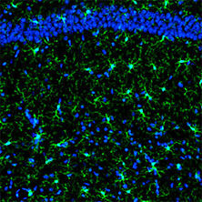
Species: Mouse
Site: Hippocampus
Sample: Frozen section
Antibody concentration: 1:1,000
Data by courtesy of
Dr. F, A University
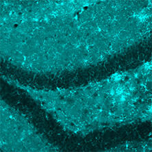
Species: Mouse (APPNL-G-F)
Site: Hippocampus
Sample: Frozen section
Antibody concentration: 1:1,000
Data by courtesy of
Dr. Takatori, Graduate School of Pharmaceutical Sciences,
University of Tokyo
Species: Mouse
Site: Brainstem (Principal sensory trigeminal nucleus)
Sample: Vibratome section
Antibody concentration: 1:500
Note: Tissue clearing with Scale method
Data by courtesy of
Dr. Ueta, School of Medicine,
Tokyo Women's Medical University
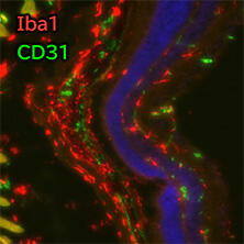
Species: Mouse
Site: Retina (Post-surgery)
Sample: Frozen section
Antibody concentration: 1:500
Data by courtesy of
Dr. Hayashi, School of Medicine, Aichi Medical University
Species: Mouse
Site: Retina (Flat-mount)
Sample: Frozen section
Antibody concentration: 1:2,000
Data by courtesy of
Dr. Watanabe, Dr. Iwagawa, University of Tokyo Hospital
Species: Mouse
Site: Retina (Flat-mount)
Antibody concentration: 1:1,000
Data by courtesy of
Dr. Watanabe, Dr. Iwagawa, University of Tokyo Hospital
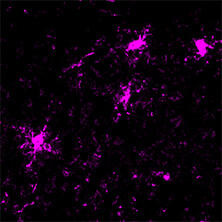
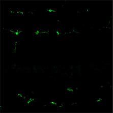
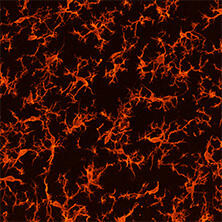
Normal
Activated
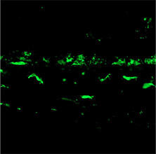
Species: Mouse
Site: Retina (Cross-section)
Sample: Frozen section
Antibody concentration: 1:2,000
Data by courtesy of
Dr. Watanabe, Dr. Iwagawa, University of Tokyo Hospital
Species: Mouse
Site: Spinal cord
Sample: Frozen section
Antibody concentration: 1:2,000
Data by courtesy of
Dr. Hagiwara and Mr. Sawada, Faculty of Science and Technology,
Tokyo University of Science
Species: Mouse
Site: Lumbar spinal cord
Sample: Frozen section
Antibody concentration: 1:1,200
Data by courtesy of
Dr. Takase, Graduate School of Medical Sciences,
Kyushu University
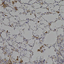
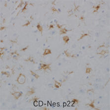
Species: Mouse
Site: Lung
Sample: Paraffin section
Antibody concentration: 1:1,000
Data by courtesy of
Dr. K, N University
Rat

Species: Rat
Site: Cerebral cortex
Sample: Frozen section
Antibody concentration: 1:1,000
Species: Rat
Site: Cerebral cortex (Medial prefrontal cortex)
Sample: Frozen section
Antibody concentration: 1:1,000
Data by courtesy of
Dr. Ohta, Faculty of Medicine, Kagawa University
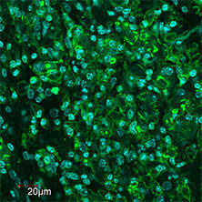
Immunocytochemistry
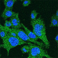
Cell line: SIM-A9
Antibody concentration: 1:200
Data by courtesy of
Dr. I, University of T
Please note that Fujifilm Wako cannot answer any questions regarding the data above.
Flow cytometry
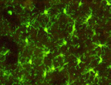
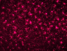
The microglial cell line BV-2 cells were fixed and permeabilized using the BD Cytofix/Cytoperm™ Fixation/Permeabilization Kit (BD Biosciences, 554714). Cells were then stained with either buffer, isotype control, or this product, followed by staining with the PE-labeled secondary antibody (SouthernBiotech, 4030-09). The fluorescence-labeled cells were analyzed by flow cytometry.
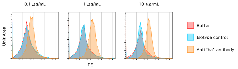
[Result]
Fixed and permeabilized BV-2 cells were successfully detected using the anti Iba1 antibody and flow cytometry.
User Voice
Researchers evaluated this antibody during the "Anti-Iba1 Rabbit Monoclonal Antibody Free Sample Campaign" held from March to May 2023 in Japan. Below are the results of the survey submitted by the researchers.
Results of the survey (N=33)
Q. Please tell us how satisfied you are with this antibody.
Q. Would you like to use this product in the future?
Q. How did this product compare to the anti-Iba1 antibody you are currently using?
References
- Imai, Y., Ibata, I., Ito, D., Ohsawa, K., & Kohsaka, S.: Biochem. Biophys. Res. Commun., 224(3), 855(1996).
A Novel Geneiba1 in the Major Histocompatibility Complex Class III Region Encoding an EF Hand Protein Expressed in a Monocytic Lineage - Mori, I., Imai, Y., Kohsaka, S., & Kimura, Y.: Microbiol. Immunol., 44(8), 729(2000).
Upregulated expression of Iba1 molecules in the central nervous system of mice in response to neurovirulent influenza A virus infection - Sasaki, Y., Ohsawa, K., Kanazawa, H., Kohsaka, S., & Imai, Y. Biochem. Biophys. Res. Commun., 286(2), 292(2001).
Iba1 is an actin-cross-linking protein in macrophages/microglia.
FAQ
About antibody
- What is the antigen?
- It is a synthetic peptide of Iba1 (homologous to the C-terminal sequence). The specific sequence is not disclosed.
About application
- Can this antibody be used for paraffin section?
- In our investigation, we have not confirmed the application of immunohistochemical staining of paraffin sections using this antibody. However, some researchers reported it can be used to immunohistochemical staining of paraffin sections. (see "application data")
- Can this antibody be used for Western blotting?
- This antibody is suitable for immunohistochemistry (frozen sections) and immunocytochemistry. For Western blotting, use Anti Iba1, Rabbit (for Western Blotting) (Product Number 016-20001).
Overview / Applications
| Outline | This product is for research use only. Do not administer it to human. Iba1 is a protein of about 17 kDa that is expressed specifically in microglia of the nervous system, and is frequently used as a microglial marker. This product is a rabbit monoclonal antibody that recognizes Iba1. It is for immunohistological staining with frozen section. [Antigen] C-terminal sequence peptide of Iba1 [Species cross reactivity] mouse, rat [Applications] Immunohistochemistry (frozen section) 1: 200-10,000 Flow CytoMetry 1:100-1:1000 [Reference] Hiramasu, K., et al.: Cell Biol. Int,. 48, 76 (2024). |
|---|
Property
| Appearance | Liquid |
|---|---|
| Concentration | Protein : 1.0 - 1.2mg/mL |
Manufacturer Information
Alias
For research use or further manufacturing use only. Not for use in diagnostic procedures.
Product content may differ from the actual image due to minor specification changes etc.
If the revision of product standards and packaging standards has been made, there is a case where the actual product specifications and images are different.