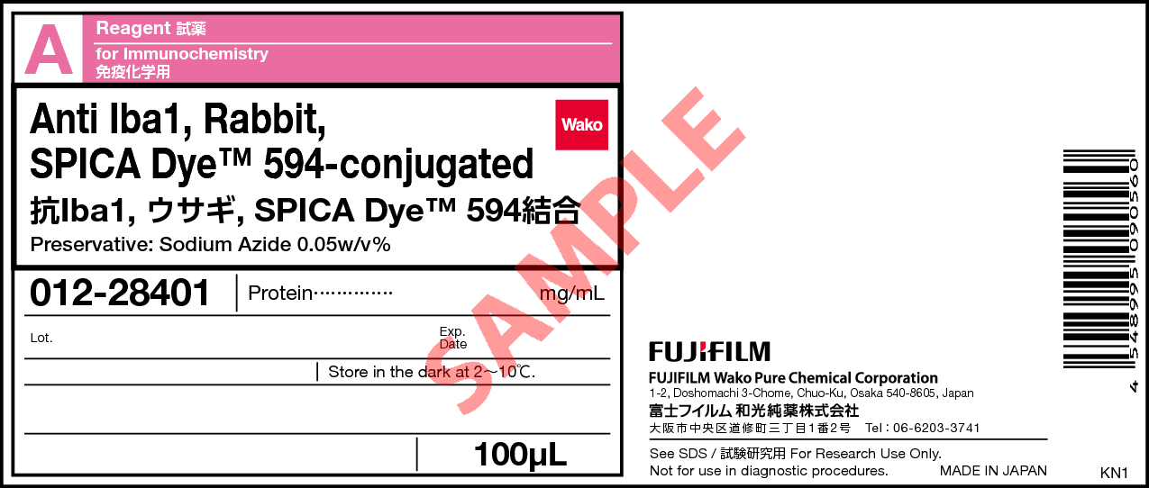
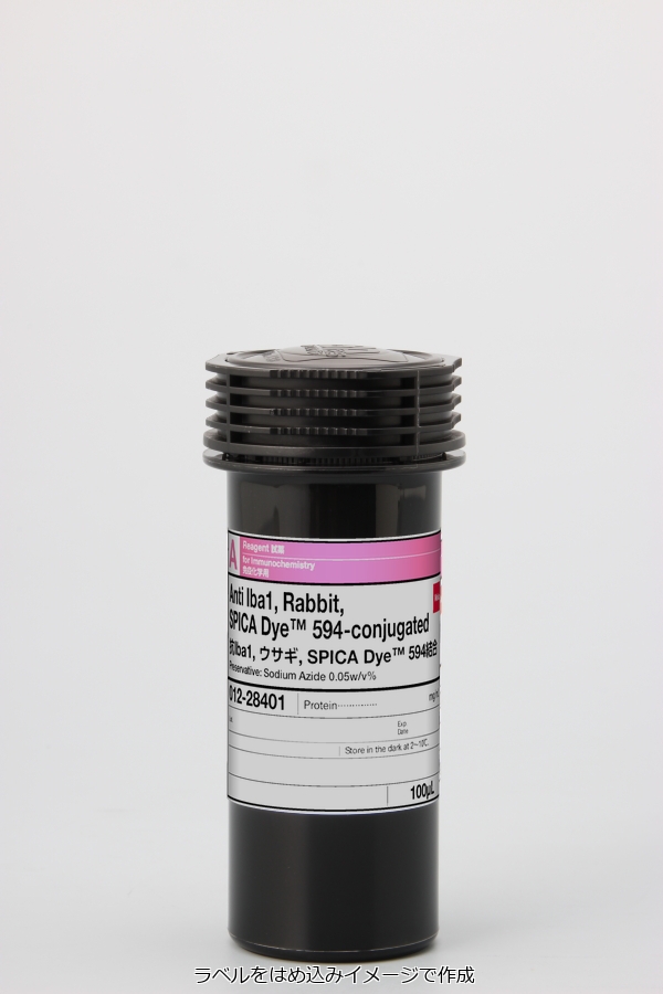
Anti Iba1, Rabbit, SPICA Dye(TM) 594-conjugated
- for Immunochemistry
- Manufacturer :
- FUJIFILM Wako Pure Chemical Corporation
- Storage Condition :
- Keep at 2-10 degrees C.
- Structural Formula
- Label
- Packing
|
Comparison
|
Product Number
|
Package Size
|
Price
|
Inventory
|
|
|---|---|---|---|---|---|
|
|
|
100uL
|
|
In stock in Japan |
Document
Overview
Iba1 (Ionized calcium-binding adapter molecule1) is an approximately 17 kDa calcium-binding protein. It is used as a microglial marker because it is expressed specifically in microglia in the central nervous system1). It is expressed in both resting and activated microglia but is reportedly expressed more highly in activated microglia2). It is also expressed in macrophages in peripheral tissues and is known as AIF-1 (Allograft inflammatory factor-1). Iba1 binds to F-actin in cells to form actin bundles. The formation of actin bundles is thought to be required for the membrane ruffling observed during cell migration and phagocytosis3).
Anti Iba1, Rabbit, SPICA Dye™ 594-conjugated is Anti Iba1, Rabbit (for Immunocytochemistry) (Product Number 019-19741) labeled with SPICA Dye™ 594, a proprietary fluorescent dye. It is possible to save time to detect with a secondary antibody.
Antibody Information
| Clonality | Polyclonal |
|---|---|
| Antigen | Synthetic peptide (Iba1 C-terminal sequence) |
| Host | Rabbit |
| Reactivity | Mouse, Rat |
| Buffer | PBS, 0.05% sodium azide |
| Conjugate | SPICA Dye™ 594 (Ex=575 nm, Em=611 nm) |
| Applications | Immunohistochemistry (Frozen Section) 1:200 - 2000 |
Application Data
Immunohistochemistry
- Rat
- Mouse
Species: Rat / Mouse
Site: Cerebral cortex
Sample: Frozen section
Antibody concentration: 1:200
References
- Imai, Y., Ibata, I., Ito, D., Ohsawa, K., & Kohsaka, S.: Biochem.Biophys. Res. Commun., 224(3), 855(1996).
A Novel Geneiba1 in the Major Histocompatibility Complex Class III Region Encoding an EF Hand Protein Expressed in a Monocytic Lineage - Mori, I., Imai, Y., Kohsaka, S., & Kimura, Y.: Microbiol. Immunol., 44(8), 729(2000).
Upregulated expression of Iba1 molecules in the central nervous system of mice in response to neurovirulent influenza A virus infection - Sasaki, Y., Ohsawa, K., Kanazawa, H., Kohsaka, S., & Imai, Y. Biochem. Biophys. Res. Commun., 286(2), 292(2001).
Iba1 is an actin-cross-linking protein in macrophages/microglia.
Overview / Applications
Property
| Appearance | Liquid |
|---|
Manufacturer Information
Alias
- Anti Iba1 Polyclonal Antibody, Rabbit, SPICA Dye (TM) 594-conjugated
For research use or further manufacturing use only. Not for use in diagnostic procedures.
Product content may differ from the actual image due to minor specification changes etc.
If the revision of product standards and packaging standards has been made, there is a case where the actual product specifications and images are different.


