Microglia Research

Microglia are a type of glial cell and are responsible mainly for the immunity of the central nervous system. Moving in the central nervous system, microglia perform various functions, including cytokine release, phagocytosis of foreign materials and dead cells, and synaptic pruning. Although microglia are essential for maintaining the function of the nervous system, their excessive activity may cause or aggravate various neurologic diseases. An association between microglia and neurological disorders such as Alzheimer's disease and amyotrophic lateral sclerosis (ALS) has been reported, and microglia have attracted attention as a key for elucidation of neurologic diseases and a target of drug discovery.
FUJIFILM Wako offers a line-up of antibodies against microglia markers, including the anti-Iba1 antibody used worldwide.
More Information
What are Microglia?
Microglia are glial cells responsible for immune function in the central nervous system. They were first described and named by the Spanish neuroscientist Pío del Río Hortega in 19191).
Unlike neurons, astrocytes, and oligodendrocytes, which are derived from the ectoderm, microglia are derived from mesodermal progenitors in the yolk sac, erythro-myeloid progenitors (EMPs)2). EMPs generated early in development migrate throughout the body, and those that migrate to the central nervous system differentiate into microglia. Later, as development progresses, EMPs are replaced in most tissues by monocyte-derived macrophages of hematopoietic stem cell (HSC) origin. However, these macrophages cannot readily penetrate the blood-brain barrier formed during development, and replacement with macrophages of HSC origin rarely occur in the central nervous system3).
Like macrophages, microglia phagocytose debris and dead cells and release chemokines and cytokines. These activities quickly remove unwanted materials such as debris and dead cells, and repair of the injured site begins following an immune response. Microglia are thus responsible for maintaining homeostasis in the central nervous system. However, with advanced neurodegenerative disease, severe neuronal injury, or chronic inflammatory responses, microglia can exacerbate the disease or injury. Microglia-targeted drug discovery not only makes use of the intrinsic function of microglia, but also attempts to block the involvement of microglia in the exacerbation of symptoms.
The morphological changes in microglia depend significantly on the environment. Normally, microglia are in a resting (ramified) form with processes extending from a relatively small cell body. However, when activated by nerve injury or other stimuli, their morphology changes to an amoeboid form with an enlarged cell body and retracted processes, resembling macrophages.
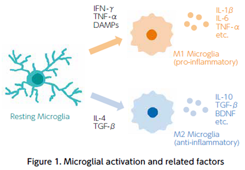
Previous studies have shown two types of activated microglia: M1 (pro-inflammatory) and M2 (anti-inflammatory) microglia4). The former responds to IFN-γ, TNF-α , and damage-associated molecular patterns (DAMPs) and releases pro-inflammatory cytokines (IL-1β , IL-6, TNF-α) and reactive oxygen species, while the latter responds to IL-4 and TGF-β and releases anti-inflammatory cytokines such as IL-10 and TGF-β, as well as trophic factors such as BDNF5) (Figure 1).
In recent years, researchers have attempted to extend the classification of microglia into subtypes. In a 2019 study, mouse microglia were analyzed by single cell RNAseq and divided into at least nine different clusters6).Microglia also play other important roles in the development and function of the central nervous system, such as extending their processes to the synapses of neurons to monitor neural status through direct contact7) and being involved in synaptic pruning8).
References
- Río-Hortega, P.d.: Biol. Soc. Esp. de Biol., 9, 68(1919).
El ‘tercer element’de los centros nerviosos. Poder fagacitario y movilidad de la microglia - Ginhoux, F., et al.: Science, 330(6005), 841(2010).
Fate mapping analysis reveals that adult microglia derive from primitive macrophages - Prinz, M., Jung, S., & Priller, J.: Cell, 179(2), 292(2019).
Microglia Biology: One Century of Evolving Concepts - Boche, D., Perry, V. H., & Nicoll, J. A. R.: Neuropath. Appl. Neurobiol., 39(1), 3(2013).
Review: activation patterns of microglia and their identification in the human brain - Bolós, M., Perea, J. R., & Avila, J.: Biomol. Concepts, 8(1), 37(2017).
Alzheimer's disease as an inflammatory disease - Hammond, T. R., et al.: Immunity, 50(1), 253(2019).
Single-Cell RNA Sequencing of Microglia throughout the Mouse Lifespan and in the Injured Brain Reveals Complex Cell-State Changes - Wake, H., et al.: J. Neurosci., 29(13), 3974(2009).
Resting microglia directly monitor the functional state of synapses in vivo and determine the fate of ischemic terminals - Schafer, D., et al.: Neuron, 74(4), 691(2012).
Microglia sculpt postnatal neural circuits in an activity and complement-dependent manner
For research use or further manufacturing use only. Not for use in diagnostic procedures.
Product content may differ from the actual image due to minor specification changes etc.
If the revision of product standards and packaging standards has been made, there is a case where the actual product specifications and images are different.