Anti Iba1 Antibodies
Iba1 (Ionized calcium-binding adapter molecule1) is used as a microglial marker because it is expressed specifically in microglia in the central nervous system. In peripheral tissues, it is expressed in macrophages and is also known as AIF-1. Fujifilm Wako's "Anti Iba1, Rabbit (for immunocytochemistry)" (Product Number 019-19741) is used by researchers all over the world as a standard for microglia marker antibody. In addition, Fujifilm Wako has also developed "Anti Iba1, Rabbit Monoclonal Antibody (6A4), recombinant" (Product Number 018-28523), which has the same performance as Anti Iba1, Rabbit (for immunocytochemistry).
About Iba1
Iba1 (Ionized calcium-binding adapter molecule1) is an approximately 17 kDa calcium-binding protein. It is used as a microglial marker because it is expressed specifically in microglia in the central nervous system1). It is expressed in both resting and activated microglia, but is reportedly expressed more highly in activated microglia2). It is also expressed in macrophages in peripheral tissues and is known as AIF-1 (Allograft inflammatory factor-1).
Iba1 binds to F-actin in cells to form actin bundles. The formation of actin bundles is thought to be required for the membrane ruffling observed during cell migration and phagocytosis3).
Anti Iba1 Antibodies of Fujifilm Wako
Fujifilm Wako’s "Anti Iba1, Rabbit (for immunocytochemistry)" (Product Number 019-19741), which allows even microglia processes to be stained by immunohistochemical staining, is used by researchers all over the world as a standard for microglia marker antibody..
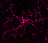
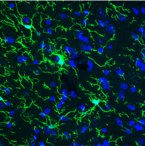
Immunohistochemical staining
of mouse cerebellum
Anti Iba1, Rabbit
(for immunocytochemistry)
Annual number of publications
Anti Iba1, Rabbit (for immunocytochemistry) has been appearing in an increasing number of publications, including many articles published in top journals (Nature, Cell, Neuron, etc.), and the annual number of such publications exceeded 1,500.
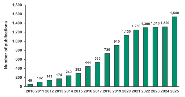
Searched in Google scholar using the keyword "Iba1 019-19741 Wako"
| Journal | Number of Publications | Impact Factor in 2024 |
|---|---|---|
| Nature | 49 | 50.5 |
| Cell | 43 | 45.5 |
| Science | 3 | 44.7 |
| Nature Medicine | 19 | 58.7 |
| Nature Neuroscience | 89 | 21.2 |
| Nature Immunology | 13 | 27.7 |
| Nature Biotechnology | 9 | 33.1 |
| Nature Methods | 4 | 36.1 |
| Nature Biomedical Engineering | 6 | 26.8 |
| Nature Cell Biology | 3 | 17.3 |
| Nature Genetics | 1 | 31.7 |
| Neuron | 67 | 14.7 |
| Total | 306 |
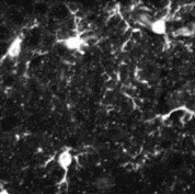
Immunohistochemical staining
of mouse cerebral cortex
Anti Iba1, Rabbit
Monoclonal Antibody (6A4), recombinant
Development of rabbit monoclonal antibody
In 2023, Fujifilm Wako launched a rabbit monoclonal antibody, "Anti Iba1, Rabbit Monoclonal Antibody (6A4), recombinant" (Product Number 018-28523). We have confirmed that this antibody has the same performance as “Anti Iba1, rabbit (for immunocytochemistry)” in immunohistological staining of mice and rat brains. It has also been reported to obtain good results in immunohistochemical staining of mouse retinas (see application data).
Product Lineup
Selection Flowchart

*2 Abbreviations:IHC(F)=Immunohistochemistry (frozen section), IHC(P)=Immunohistochemistry (paraffin section), ICC=Immunocytochemistry; WB=Western blotting
Product Lineup
| Host | Rabbit | Goat | Mouse | ||||||||
|---|---|---|---|---|---|---|---|---|---|---|---|
| Product Number | 018-28523 | 019-19741 | 013-27691 | 016-20001 | 016-26461 | 015-28011 | 012-28401 | 013-26471 | 011-27991 | 016-26721 | 013-27593 |
| Description | Rabbit monoclonal antibody | For Immno- staining (Standard) | For paraffin section | For western blotting | Biotin-conjugated | SPICA Dye™ 568-conjugated | SPICA Dye™ 594-conjugated | Red fluorochrome (635)-conjugated | Goat polyclonal antibody | Mouse monoclonal antibody (NCNP24) | Mouse monoclonal antibody (NCNP27) |
| Conjugate | Unconjugated | Unconjugated | Unconjugated | Unconjugated | Biotin | SPICA Dye™ 568 Ex=556 nm Em=591 nm |
SPICA Dye™ 594 Ex=575 nm Em=611 nm |
Red fluorochrome (635) Ex=634 nm Em=654 nm |
Unconjugated | Unconjugated | Unconjugated |
| Antibody | Monoclonal antibody | Polyclonal antibody | Monoclonal antibody | ||||||||
| Concentration (mg/mL) | 1.0-1.2 | 0.5-0.7 | 0.5-0.7 | 0.5-0.7 | 0.5-0.6 | 0.5-0.6 | 0.5-0.6 | 0.5-0.6 | 0.6-0.7 | 0.9-1.6 | 0.9-1.3 |
| Antigen | Synthetic peptide (Iba1 C-terminal sequence) | ||||||||||
| Cross-reactivity | Mouse Rat |
Mouse Rat Other species also reported*1 |
Mouse Rat |
Human Mouse Rat |
Marmoset Mouse Rat |
Mouse Rat |
Mouse Rat |
Mouse Rat |
Mouse Rat |
Marmoset Mouse Rat |
Human |
| Application*2 Figures indicate recommended concentrations | IHC(F) 1:200-10,000 FCM 1:100-10,000 |
IHC(F) 1:500-1,000 ICC 1:500-1,000 |
IHC(P) 1:500-1,000 |
WB 1:500-1,000 |
IHC(F) 1:200-2,000 |
IHC(F) 1:200-2,000 |
IHC(F) 1:200-2,000 |
IHC(F) 1:200-2,000 |
IHC(F) 1:250-1,000 IHC(P) 1:250-1,000 WB 1:1,000 |
IHC(F, DAB) 1:500-2,000 IHC(F, fluorescent) 1:100 |
IHC(P, DAB) 1:100-1,000 |
| Volume | 100 μL | 50 μg | 50 μg | 50 μg | 100 μL | 100 μL | 100 μL | 100 μL | 100 μL | 50 μL | 50 μL |
*2 Abbreviations:IHC(F)=Immunohistochemistry (frozen section), IHC(P)=Immunohistochemistry (paraffin section), ICC=Immunocytochemistry; WB=Western blotting
Standard Protocol for Immunohistochemistry of Microglia using Iba1 antibody
Fujifilm Wako's "Anti Iba1, Rabbit (for Immunocytochemistry)" (Product No. 019-19741) is an excellent microglial marker antibody since it can stain even microglial processes. Here, we describes the protocol and points to note when performing microglial immunohistochemistry. Frozen sections of mouse brain and a fluorescent dye are used as an example.
1.Preparation of tissue sections
1-1. Mice are perfused and fixed in 4% paraformaldehyde-phosphate buffer.
1-2. Replace with sucrose, and prepare frozen blocks.
1-3. Prepare 50 µm-sections in thickness using a microtome.
2.Washing - Blocking
2-1. Wash with 0.3% TritonX-100/PBS 3 times for 5 minutes each.
2-2. Block in 1% BSA, 0.3% TritonX-100/PBS for 2 hours at room temperature.
The following blocking solutions can also be used:
· 1% BSA, 0.3% Tween-20/PBS
· 3% normal serum of the host of the secondary antibody
3.Primary antibody reaction
3-1. Add Anti Iba1, Rabbit (for immunohistochemistry) to 1% BSA, 0.3% TritonX-100/PBS at 1:1,000 dilution.
3-2. Incubate overnight at 4°C.
4.Washing
4-1. Wash with 0.3% TritonX-100/PBS 3 times for 5 minutes each.
5.Secondary antibody reaction
5-1. Add fluorescent-labeled anti-rabbit IgG antibody (e.g. Jackson ImmunoResearch, Product No. 111-545-144), to 1% BSA, 0.3% TritonX-100/PBS at 1:1,000 dilution.
5-2. Incubate for 2 hours at room temperature.
6.Washing
6-1. Wash with 0.3% TritonX-100/PBS 3 times for 5 minutes each.
7.Mounting
7-1. Mount the sections in mounting media.
8.Observation
8-1. Observe the sections under a fluorescence microscope or a confocal microscope.
If microglia do not stain well using the techniques described at left, perform antigen retrieval after sectioning using one of the following:
(A) Citrate buffer (pH 6.0) for 9 minutes at 90°C
(B) TE bufferi (pH 9.0) for 9 minutes at 90°C
Other troubleshooting is also available in the FAQ on the product detail page.
Application Data
Immunohistochemistry
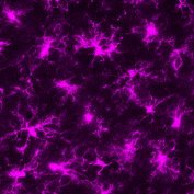
▼Anti Iba1, Rabbit Monoclonal Antibody (6A4), recombinant
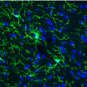
Species: Mouse
Site: Cerebral cortex
Sample: Frozen section
Antibody concentration: 1:1,000
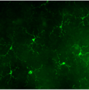
Species: Mouse
Site: Retina
Sample: Frozen section
Antibody concentration: 1:2,000
Data by courtesy of
Dr. Watanabe, Dr. Iwagawa
University of Tokyo Hospital
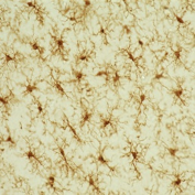
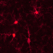
▼Anti Iba1, Rabbit (for immunocytochemistry)
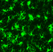
Species: Rat
Site: Cerebral cortex
Sample: Frozen section
Antibody concentration: 1:1,000
Data by courtesy of
Dr. Sanagi, Dr. Manabe,
Dr. Ichinohe, Dr. Kohsaka
National Center of
Neurology and Psychiatry
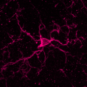
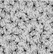
Species: Mouse
Site: Cerebellum
Sample: Frozen section
Antibody concentration: 1:1,000

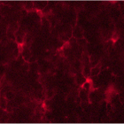
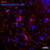
- Species
- Mouse
- Site
- Cerebral cortex/Area postrema (Medulla oblongata)
- Sample
- Frozen section
- Antibody concentration
- P2RY12 → Anti P2RY12, Guinea Pig (Product No. 011-28873) 1:900
Iba1 → Anti Iba1, Rabbit (for Immunocytochemistry) 1:500
Laminin → Anti Laminin Antibody (Rat, Made in Dr. Miyata’s Lab) 1:200
Data by courtesy of
Dr. Miyata, Department of Applied Biology, Kyoto Institute of Technology
[Result]
Among the Iba1-positive cells, those that are present in the brain parenchyma are P2RY12-positive and are thought to be microglia (arrowheads). On the other hand, Iba1-positive/P2RY12-negative cells are present around the blood vessel basement membrane, and it is thought that these cells are macrophages (arrows).
▼Anti Iba1, Rabbit (for paraffin section)
Species: Rat
Site: Hippocampus vicinity
Sample: Paraffin section
Antibody concentration: 1:1,000
▼Anti Iba1, Rabbit, Biotin-conjugated
Species: Rat
Site: Cerebral cortex
Sample: Frozen section
Antibody concentration: 1:200
Data by courtesy of
Dr. Sanagi, Dr. Manabe,
Dr. Ichinohe, Dr. Kohsaka
National Center of
Neurology and Psychiatry
▼Anti Iba1, Rabbit, SPICA Dye™ 568-conjugated
Species: Rat
Site: Cerebral cortex
Sample: Frozen section
Antibody concentration: 1:200
▼Anti Iba1, Rabbit, SPICA Dye™ 594-conjugated
Species: Rat
Site: Cerebral cortex
Sample: Frozen section
Antibody concentration: 1:200
▼Anti Iba1, Rabbit, Red Fluorochrome (635)-conjugated
Species: Rat
Site: Cerebral cortex
Sample: Frozen section
Antibody concentration: 1:200
Data by courtesy of
Dr. Sanagi, Dr. Manabe,
Dr. Ichinohe, Dr. Kohsaka
National Center of
Neurology and Psychiatry
Species: Mouse
Site: Hippocampus
Antibody concentration: 1:200
Data by courtesy of
Dr. Takata
Division of Integrated
Pharmaceutical Sciences,
Kyoto Pharmaceutical University
▼Anti Iba1, Mouse Monoclonal Antibody (NCNP24)
Species: Rat
Site: Cerebral cortex
Sample: Frozen section
Antibody concentration: 1:1,000
Data by courtesy of
Dr. Sanagi, Dr. Manabe,
Dr. Ichinohe, Dr. Kohsaka
National Center of
Neurology and Psychiatry
Western blotting
▼Anti Iba1, Rabbit (for Western Blotting)
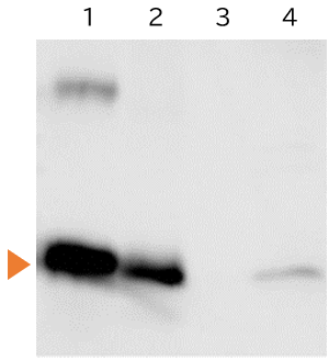
1.Iba1 protein...10 ng
2.Rat Microglia...10 μg
3.Rat Neuron...10 μg
4.Rat cerebral cortex...150 μg
SDS-PAGE: 5.5% stacking gel, 12.5% running gel, 100V
Blocking: 3% skim milk/TBS, 1 hour, room temperature.
Primary antibody: 1:1,000 concentration, 3% skim milk/TTBS, overnight, 4℃
Secondary antibody: Peroxidase-labeled Anti Rabbit IgG (1/5,000), 3% skim milk/TTBS, 1 hour, room temperature
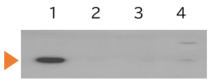
1.Rat primary culture microglia...10 μg
2.Rat primary culture neuron...10 μg
3.Rat primary culture astrocyte...10 μg
4.Rate cerebral cortex...100 μg
Primary antibody: Anti Iba1, Goat, 1:1,000
Secondary antibody: Anti Goat IgG, HRP-conjugated
References
- Imai, Y., Ibata, I., Ito, D., Ohsawa, K. & Kohsaka, S.: Biochemical and biophysical research communications, 224(3), 855(1996).
A Novel Geneiba1 in the Major Histocompatibility Complex Class III Region Encoding an EF Hand Protein Expressed in a Monocytic Lineage - Mori, I., Imai, Y., Kohsaka, S. & Kimura, Y.: Microbiology and immunology, 44(8), 729(2000).
Upregulated expression of Iba1 molecules in the central nervous system of mice in response to neurovirulent influenza A virus infection - Sasaki, Y., Ohsawa, K., Kanazawa, H., Kohsaka, S. & Imai, Y.: Biochemical and biophysical research communications, 286(2), 292(2001).
Iba1 is an actin-cross-linking protein in macrophages/microglia. - Zhao, S. et al.: Cell, 180(4), 796(2020).
Cellular and Molecular Probing of Intact Human Organs - Ahn, J.H. et al.: Lab. Anim. Res., 28(3), 165 (2012).
Comparison of alpha-synuclein immunoreactivity in the spinal cord between the adult and aged beagle dog - Ide, T. et al.: J. Vet. Med .Sci., 72(1), 99 (2010).
Histiocytic Sarcoma in the Brain of a Cat - Gaige, S. et al.: Neurotoxicology, 34, 135(2013).
c-Fos immunoreactivity in the pig brain following deoxynivalenol intoxication: Focus on NUCB2/nesfatin-1 expressing neurons - Rodriguez-Callejas, J.D. et al.: Front. Aging Neurosci., 8, 315(2016).
Evidence of Tau Hyperphosphorylation and Dystrophic Microglia in the Common Marmoset - Fantin, A. et al.: Blood, 116(5), 829(2010).
Tissue macrophages act as cellular chaperones for vascular anastomosis downstream of VEGF-mediated endothelial tip cell induction
Product List
- Open All
- Close All
Anti Iba1, Rabbit (Monoclonal Antibody)
Anti Iba1, Rabbit (Polyclonal Antibody)
Anti Iba1, Goat (Polyclonal Antibody)
Anti Iba1, Mouse (Monoclonal Antibody)
For research use or further manufacturing use only. Not for use in diagnostic procedures.
Product content may differ from the actual image due to minor specification changes etc.
If the revision of product standards and packaging standards has been made, there is a case where the actual product specifications and images are different.