Anti CX3CR1 Antibodies
CX3C motif chemokine receptor 1 (CX3CR1) is a receptor for CX3CL1, also known as fractalkine, and belongs to the G protein-coupled receptor (GPCR) family. In peripheral tissues, CX3CR1 is primarily expressed on the surface of macrophages, lymphocytes, and dendritic cells, and it participates in processes such as cell survival, proliferation, adhesion, and migration. In the central nervous system (CNS), CX3CR1 is predominantly expressed in microglia and macrophages, playing a role in the development and maintenance of homeostasis within the CNS.
What is CX3CR1?
CX3CR1 is a receptor for CX3CL1, also known as fractalkine, and belongs to the GPCR family. In peripheral tissues, CX3CR1 is primarily expressed on the surface of macrophages, lymphocytes, and dendritic cells, and it participates in processes such as cell survival, proliferation, adhesion, and migration.
In the CNS, CX3CR1 is predominantly expressed in microglia and macrophages, serving as a marker for these cells. It plays a role in the development and maintenance of homeostasis within the CNS1). Notably, CX3CR1 knockout mice exhibit deficits in synaptic pruning by microglia, leading to abnormalities in synaptic transmission between neurons. This suggests that CX3CR1 is critical for synaptic pruning2).
Anti CX3CR1, Guinea Pig
Anti CX3CR1, Guinea Pig is a polyclonal antibody against CX3CR1, derived from guinea pigs3-4). It is suitable for use in multiplex immunohistochemical staining.
Antibody Information
| Clonality | Polyclonal |
|---|---|
| Antigen | CX3CR1 synthetic peptide (corresponding to N-terminal sequence) |
| Host | Guinea Pig |
| Formulation | PBS, 0.1% sodium azide |
| Conjugate | Unconjugated |
| Cross-reactivity | Mouse, Rat |
| Application | IHC (frozen section) 1:800-1,600 |
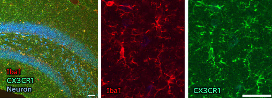
Application Data
Immunohistochemistry

- Species
- Mouse
- Site
- Hippocampus
- Sample
- Frozen section
- Primary antibody
- Anti Iba1, Rabbit (for Immunocytochemistry)
(Product Number 019-19741) 1:500
Anti CX3CR1, Guinea Pig (this product) 1:800
NeuroTrace™435/455 Blue Fluorescent Nissl Stain (ThermoFisher Scientific, Inc.) was used for staining neurocytes.
[Data by courtesy of]
Dr. Miyata, Department of Applied Biology, Kyoto Institute of Technology
References
- Wolf, Y. et al.: Front. Cell. Neurosci., 7, 26(2013).
Microglia, seen from the CX3CR1 angle - Zhan, Y. et al.: Nat. Neurosci., 17(3), 400(2014).
Deficient neuron-microglia signaling results in impaired functional brain connectivity and social behavior - Takagi, S. et al.: J. Neuroimmunology, 331, 74(2019).
Microglia are continuously activated in the circumventricular organs of mouse brain - Kawai, S. et al.: Cell Biochem. Funct., 38(4), 392(2020).
Transient increase of microglial C1q expression in the circumventricular organs of adult mouse during LPS‐induced inflammation
Product List
- Open All
- Close All
Anti CX3CR1, Guinea Pig
For research use or further manufacturing use only. Not for use in diagnostic procedures.
Product content may differ from the actual image due to minor specification changes etc.
If the revision of product standards and packaging standards has been made, there is a case where the actual product specifications and images are different.


