



Brain Organoids / Neural Organoids
Brain organoids and neural organoids are more than cell aggregates consisting of a single cell type induced from ES or iPS cells; they are three-dimensional tissue structures that contain multiple cell types and partially recapitulate the architecture of the brain and neural tissue. The ability to observe the process of human brain formation in vitro has opened the way to research on neurogenesis in the human fetal period and on pathological conditions such as disease-specific signaling abnormalities. This section introduces the fundamentals of brain organoids and neural organoids, including the principles underlying their generation, differentiation induction protocols, and the reagents used for their production.

Brain Organoids
The human brain has an extremely complex structure, with each region responsible for its own distinct functions. Recent advances in stem cell technology have made it possible to generate brain region-specific organoids that recapitulate the characteristics of each region. These organoids are helping to elucidate human brain development, clarify the pathology of intractable psychiatric and neurological disorders, and drive drug discovery research.
Cerebral Organoids
The cerebrum accounts for more than half of the brain's volume and serves as the command center underlying complex phenomena such as perception, thought, language, attention, episodic memory, and voluntary movement. Cerebral organoids are emerging as a promising model for studying aspects of human brain development that remain poorly understood. A key example is the diversification of human cortical cell populations—such as outer radial glia (oRG) in the upper cortical layers and callosal projection neurons (CPN)—which underwent marked, species-specific expansion during human evolution.
Development and Structure of the Cerebral Cortex in vivo
The cerebral cortex is formed through the following developmental process:
- Early proliferative phase (neural epithelium proliferation)
After neural tube closure, neural epithelium cells (NECs) repeatedly self-renew through symmetric division, expanding the neuroepithelial cell pool. - Neurogenesis (neuron supply)
With the initiation of neurogenesis, NECs transition into radial glial cells (RGCs). RGCs whose cell bodies lie on the ventricular side, with processes extending toward the basal surface, are specifically referred to as apical radial glial cells (aRGCs). These cells undergo asymmetric division, producing one aRGC and either an intermediate progenitor cell (IPC) or a neuron. - Formation of the six-layer structure
Neurons generated by aRGCs are sequentially arranged from the deep layers (layers V/VI) to the superficial layers (layers II/III/IV), giving rise to the distinct six-layered structure of the cerebral cortex. - Regulation by the marginal zone (MZ)
Cajal–Retzius cells, which originate from regions outside the cerebral cortical primordium, migrate tangentially to form the marginal zone (MZ), where they regulate the proper laminar positioning of neurons.
Interspecies Differences in the Cerebral Cortex
The number of neurons in the human cerebral cortex reaches approximately 16 billion—a staggering scale that is about 1,000 times that of mice and roughly 2.7 times that of chimpanzees (approximately 6 billion), our closest living relatives. This cell number is directly influenced by the size of the neuroepithelial cell (NEC) pool during early development. The longer the NEC proliferative period, the greater the final neuron count, and this proliferative period has indeed been confirmed to be longer in primates than in rodents.
A notable feature of the human cerebral cortex is the markedly higher proportion of pyramidal neurons located in layers II/III (the upper cortical layers). Whereas upper-layer cortical neurons in mice consist of three subtypes, humans have five subtypes. Because each layer of the cerebral cortex has its own distinct function, this diversity of upper-layer neurons (the acquisition of diverse subtypes) is thought to underlie the advanced cognitive functions unique to humans and the evolution of higher brain function.
Another important distinction is the presence or absence of the folded structure (gyrification) of the cerebral cortex. This structure allowed humans to expand the cortical surface area within a limited cranial volume. The outer subventricular zone (oSVZ) and basal radial glial cells (bRGCs) are abundant in gyrencephalic mammals such as humans but are nearly absent in mice. This suggests that bRGCs are an important source of neural progenitor cells contributing to cortical expansion and folding and play a critical role in the formation of gyri and sulci in the human brain.
Cerebral Cortex Organoid Induction Protocols
Methods for inducing cerebral cortex organoids from human pluripotent stem cells (hESCs/hiPSCs) are classified into two approaches: pre-patterning and self-patterning.
1. Pre-patterning
This method involves the addition of exogenous signals (small-molecule compounds or growth factors), to differentiation induction toward specific target brain regions.
Conventional directed neural differentiation from mouse ES cells mainly relied on embryoid body (EB) formation under serum-containing conditions. However, its low neural differentiation efficiency remained a persistent bottleneck. To address this, Sasai, Watanabe and colleagues developed the SFEB method, a floating culture technique using serum-free medium. They succeeded in inducing Foxg1, a transcription factor specifically expressed in the telencephalon, ventricular zone, subventricular zone, and intermediate zone, confirming induction of telencephalic progenitor cells. Further, introducing a ROCK inhibitor into the culture system greatly improved cell survival, making it possible to reproduce these results in human cells.
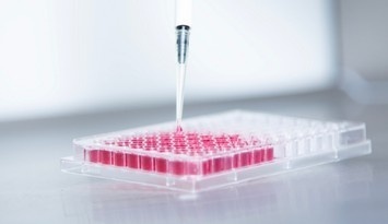
The SFEB method, however, had reproducibility issues, including variability in the size of the resulting cell aggregates and induction efficiencies for Foxg1-positive cells that fell below 50%. Eiraku and colleagues resolved this issue with the SFEBq method. By using low-adhesion 96-well plates to promote rapid cell aggregation, they succeeded in producing uniform aggregates. As a result, the efficiency of Foxg1 expression was increased to a maximum of 75%.
2. Self-patterning
This approach maximally exploits the stem cells' inherent capacity for self-organization, without relying on exogenous patterning factors.
The protocol developed by Lancaster and colleagues promotes the stem cells' intrinsic differentiation potential (self-patterning) without using exogenous signaling factors. Instead of controlling differentiation with compounds, EBs are embedded in Matrigel droplets and cultured in a spinning bioreactor, which enhances nutrient absorption and three-dimensional cell growth.
In this culture system, a neuroepithelial sheet exhibiting apical N-cadherin polarity forms around ventricle-like cavities. Long-term culture of more than 10 months is possible, and structures resembling choroid tissue and retina form alongside cerebral cortical tissue. Forebrain, midbrain, and hindbrain markers are initially detected, but forebrain marker expression becomes predominant and is maintained as culture progresses.
Despite the absence of exogenous patterning factors, expression of the forebrain marker Foxg1 and the cortical marker Emx1 is ultimately detected. Furthermore, layered structures resembling the ventricular zone, subventricular zone, and cortical plate, as well as structures and marker expression characteristic of bRGCs—primate-specific neural stem cells—have been observed within the tissue.
Midbrain Organoid
The midbrain is a complex brain region consisting of nuclei, nerves, and neural pathways involved in vision, sensation, cognitive function, and motor control. In particular, the dopaminergic nuclei located ventral to the cerebral aqueduct—namely the ventral tegmental area (VTA) and substantia nigra—play a critical role in reward, cognition, and voluntary movement control. Because dysfunction of the midbrain dopamine (mDA) system has been linked to a wide range of neurological and neuropsychiatric disorders—including Parkinson's disease (PD), attention deficit hyperactivity disorder, autism, drug addiction, and pediatric neurological disorders—midbrain organoids are useful as a research model that recapitulates important spatial and molecular features of these diseases.
Cerebellar Organoids
In addition to its role in motor behavior, the cerebellum has recently attracted attention for its involvement in cognitive function such as language, spatial cognition, working memory, and emotional processing. The cerebellum is closely associated with the pathogenesis of several disorders, including Dandy-Walker malformation, cerebellar vermis hypoplasia, and medulloblastoma. Research into hPSC-derived cerebellar organoids is advancing to elucidate the mechanisms of these diseases and to support drug discovery and treatment strategies.
Hypothalamic Organoids
The hypothalamus is responsible for maintaining homeostasis, including the regulation of body temperature, circadian rhythm, feeding, and reproduction. It plays a critical role in linking the nervous and endocrine systems by working with the pituitary gland to regulate hormone secretion. Beyond metabolic disorders (obesity, diabetes, diabetes insipidus) and sleep disorders, hypothalamic dysfunction is also implicated in neurodegenerative diseases such as Alzheimer's disease and Parkinson's disease. In recent years, hPSC-derived organoids have emerged as a promising model for these hypothalamic disorders.
Striatal Organoids
The striatum helps connect neural circuits in the human brain, and its dysfunction leads to neurological disorders such as Huntington’s disease (HD). Striatal organoids provide a useful platform for studying striatal development and associated diseases, mapping interregional neural circuits, and testing therapeutic strategies.
Protocol for the Generation of Brain Organoids
We present protocols and recommended reagents for generating cerebral, midbrain, cerebellar, hypothalamic, and striatal organoids.
Protocol for the Generation of Cerebral Organoids
These are the reagents used in each step of cerebral organoids generation. Please refer to the references for detailed protocol information. (Supakul, S., et al., 2023)
| iPSCs | Cell Aggregates | Cortical Brain Organoids | |||||
|---|---|---|---|---|---|---|---|
 |
|||||||
| Media | hPSC Medium | DMEM/F12 | DMEM/F12 | DMEM/F12 | DMEM/F12 | ||
| Serum/Additives | PS | SSR/NEAA/2-ME/ L-Ala-L-Gln/PS | NEAA/L-Ala-L-Gln/N2/PS | N2/B27-VitaminA/NEAA/ 2-ME /Insulin/L-Ala-L-Gln/PS/CDLC | N2/B27/NEAA/2-ME/Insulin/L-Ala-L-Gln/PS/CDLC | ||
| Scaffolding Materials | iMatrix-511Silk | EHS gel matrix | |||||
| Small Molecules | SB431542/IWP-2/Y-27632 | SB431542/CHIR99021 | |||||
| LDN193189 | |||||||
| Equipmen | 96-well plate | ||||||
*Created based on data from Supakul, S., et al., 2023
Protocol for the Generation of Midbrain Organoids
These are the reagents used in each step of midbrain organoid generation. Please refer to the references for detailed protocol information. (X, Yao et al., 2023)
| iPSCs | Embryoid Body |
Neuroectoderm | Mesencephalic Floor Plate | Midbrain Organoids | |||
|---|---|---|---|---|---|---|---|
 |
|||||||
| Media | hPSC Medium | DMEM/F12 | DMEM/F12/Neurobasal medium | DMEM/F12/Neurobasal medium | |||
| Serum/Additives | SSR/FBS/NEAA/2-ME/L-Ala-L-Gln/PS/Heparin | N2/B27-VitaminA/L-Ala-L-Gln/PS/NEAA/2-ME/Heparin | N2/B27/L-Ala-L-Gln/PS/NEAA/2-ME/Heparin/L-Ascorbic Acid/cAMP | ||||
| Laminin/Insulin | |||||||
| Cell Dissociation Reagents | Trypsin-EDTA | ||||||
| Scaffolding Materials | EHS gel matrix | ||||||
| Small Molecules | Y-27632 | Dordsomorphin/A83-01/IWP-2/CHIR99021 | SAG | ||||
| Cytokine | bFGF | bFGF | FGF8 | ||||
| BDNF/GDNF | |||||||
| Equipmen | 96-well plate | ||||||
*Created based on data from X, Yao et al., 2023
Protocol for the Generation of Cerebellar Organoids
These are the reagents used in each step of cerebellar organoid generation.Please refer to the references for detailed protocol information. (Atamian, A., et al., 2024)
| iPSCs | MidBrain/Rhombencephalon | Ventricular Zone、Rhombic Lip | ||||||
|---|---|---|---|---|---|---|---|---|
 |
||||||||
| Media | hPSC medium | IMDM/F12 | DMEM/F12 | DMEM/F12/Neurobasal medium | ||||
| Serum/Additives | PS | Monothioglycerol/BSA/Transferrin(Apo)/insulin/CDLC | SSR/Transferrin(Apo)/insulin/PS/L-Ala-L-Gln/2-ME | N2/B27/ L-Ala-L-Gln /PS | N2/B27/ L-Ala-L-Gln/PS/Heparin/Amphotericin B | |||
|
Cell Dissociation Reagents |
Accutase | |||||||
| Scaffolding Materials | EHS gel matrix | EHS gel matrix | ||||||
| Small Molecules | SB431542/CHIR99021 | |||||||
| Y-27632 | T3 | |||||||
| Cytokine | bFGF | Noggin | ||||||
| FGF8b | BDNF | |||||||
| Equipmen | 96-well plate | 10-cm culture plates | ||||||
*Created based on data from Atamian, A., et al., 2024
Protocol for the Generation of Hypothalamic Organoids
These are the reagents used in each step of hypothalamic organoid generation.Please refer to the references for detailed protocol information. (Sarrafha, L., et al., 2023)
| iPSCs | Hypothalamic-pattern | Hypothalamic Organoids | |||||
|---|---|---|---|---|---|---|---|
 |
|||||||
| Media | hPSC medium | DMEM/F12 | |||||
| Serum/Additives | PS | PS/L-Ala-L-Gln/B27-VitaminA/N2 | |||||
| dcAMP | |||||||
| Cell Dissociation Reagents | Accutase | ||||||
| Scaffolding Materials | EHS gel matrix | ||||||
| Small Molecules | Y-27632 | LDN193189/SB431542/XAV939 | |||||
| Thiazovivin | Purmorphamine/SAG | DAPT | |||||
| Cytokine | bFGF | BDNF/GDNF | |||||
*Created based on data from Sarrafha, L., et al., 2023
Protocol for the Generation of Striatal Organoids
These are the reagents used in each step of striatal organoid preparation.Please refer to the references for detailed protocol information. (X, Chen et al., 2022)
| iPSCs | Lateral Ganglionic Eminence | Striatal Organoids | |||||
|---|---|---|---|---|---|---|---|
 |
|||||||
| Media | DMEM/F12/hPSC medium | DMEM/F12/Neurobasal media | |||||
| Serum/Additives | NEAA/L-Ala-L-Gln/N2 | L-Ala-L-Gln/N2/B27/PS/NEAA | |||||
| Cell Dissociation Reagents | Accutase | ||||||
| Scaffolding Materials | EHS gel matrix | ||||||
| Small Molecules | Y-27632 | ||||||
| LDN193189/SB431542 | Purmorphamine | ||||||
| Cytokine | BDNF/GDNF | ||||||
| Equipmen | 96-well plate | 60mm culture plates | |||||
*Created based on data from X, Chen et al., 2022
Neural Organoids
Human stem cell-derived neurons provide an important foundation for drug discovery and the development of treatments for serious neurological diseases. They are also widely studied to understand neural development, function, and dysfunction. In recent years, appropriate multicellular tissue and microenvironments have proven essential for physiological modeling. As a result, in vitro three-dimensional organoid culture systems, which more accurately recapitulate multicellular structures, are increasingly being adopted.
Regionalized Organoids
Regionalized organoids refer to the technology—and the resulting tissue—for creating specific functional and structural regions (patterning) within a single organoid by recapitulating in vivo developmental processes when generating three-dimensional organs or tissues from pluripotent stem cells (such as iPS cells).
Examples of Regionalized Organoids
In addition to the organoids introduced above, many other methods for generating organoids have been reported, such as those described below.
1. Choroid Plexus Organoids
This organoid originates from the roof plate, located at the most dorsal part of the dorsomedial telencephalic region, and forms in a region where high concentrations of BMP and Wnt signals are present during development. Based on cerebral cortex induction conditions, a canonical Wnt signaling activator (CHIR99021) and BMP4 protein are continuously added from culture day 18 onward, once neural regionalization is complete. This enables the efficient three-dimensional induction of choroid plexus epithelial tissue that is positive for LMX1A, OTX2, and transthyretin.
2. Hippocampus Organoids
This organoid originates from the medial pallium of the medial telencephalon and is positioned between the cerebral cortex and the choroid plexus. As with choroid plexus induction, CHIR99021 and BMP4 are used, but only for three days, from culture day 18 to 21. This induces the dorsomedial telencephalon. Long-term culture (60 days or more) leads to the expression of the hippocampal marker ZBTB20 and the dentate gyrus neuron marker PROX1.
3. Spinal Cord Organoids
During spinal cord development, concentration gradients of factors secreted by organizers (dorsal BMP/Wnt and ventral Shh signaling) pattern the dorsal region into six progenitor domains and the ventral region into five.
Deliberately removing the BMP inhibitor (LDN-193189) and Shh agonist (SAG) from the conventional motor neuron induction protocol induces a broader, continuous neuroepithelium spanning from the dorsal to the intermediate regions. Concentration-dependent addition of exogenous SAG then enables precise control of patterning toward the intermediate or ventral spinal cord.
4. Ventral Telencephalon Organoids
This region primarily generates inhibitory interneurons and comprises the lateral ganglionic eminence (LGE), medial ganglionic eminence (MGE), and caudal ganglionic eminence (CGE).
Shh signaling drives patterning into the LGE (GSH2-positive), MGE (NKX2.1-positive), and CGE (CoupTFII-positive). Under cerebral cortex differentiation conditions, adding a low concentration (30 nM) of SAG during culture days 15–21 gives rise to PAX6-positive (cerebral cortex) and GSH2-positive (LGE) regions within a continuous epithelium. In contrast, adding a high concentration (500 nM) of SAG suppresses PAX6 and GSH2 expression, efficiently inducing an NKX2.1-positive neuroepithelium and achieving complete ventralization.
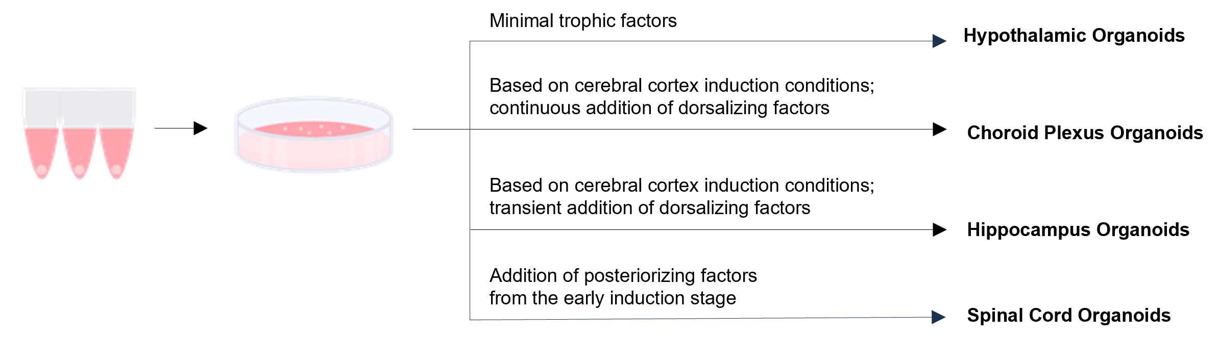
Regionalization of Neural Organoids through Controlled Culture Conditions
References
- A, Uzquiano et al.: Cell., 185, 3770 (2022)
- T, Kitahara et al.: Stem Cell Reports., 15, 467 (2020)
- D, Budinger et al.: Nat Commun., 17, 1354 (2026)
- J, Kawada et al.: Stem Cell Reports., 9, 1441 (2017)
- R, Hagiwara、M, Eiraku:Organoid research, 133 (2024).
- H, Sakaguchi:Organoid research, 125 (2024).
For research use or further manufacturing use only. Not for use in diagnostic procedures.
Product content may differ from the actual image due to minor specification changes etc.
If the revision of product standards and packaging standards has been made, there is a case where the actual product specifications and images are different.
The prices are list prices in Japan.Please contact your local distributor for your retail price in your region.