PS Capture(TM) Exosome Flow Cytometry Kit
- for Genetic Research
- Manufacturer :
- FUJIFILM Wako Pure Chemical Corporation
- Storage Condition :
- Keep at -20 degrees C.
- GHS :
-


-
 Close
Close -
 Close
Close
- Structural Formula
- Label
- Packing
- SDS
|
Comparison
|
Product Number
|
Package Size
|
Price
|
Availability
|
Certificate of Analysis
|
Purchase |
|---|---|---|---|---|---|---|
|
|
|
300Tests
|
|
In stock in Japan |
※Check availability in the US with the distributor.
Document
Kit component
Kit Components (300 assays)
| Exosome Capture Beads | 3 mL x 1 |
|---|---|
| Washing Buffer (10×) | 45 mL x 2 |
| Exosome Binding Enhancer (100×) | 15 mL x 2 |
Product Overview
PS Capture™ Exosome Flow Cytometry Kit is designed for highly sensitive detection of EVs, including exosomes, with maker proteins of interest by flow cytometry. It immobilizes EVs on magnetic beads using the PS affinity method, which utilizes phosphatidylserine-specific binding protein Tim4 and magnetic beads.
[Note]
A fluorescent-labeled primary antibody against marker protein, or a primary antibody and a fluorescent-labeled secondary antibody need to be supplied.
Features
- Highly sensitive qualitative analysis by flow cytometry
- Easy operation with magnetic beads
- No purification necessary; samples can be directly measured
- Assay completes in 3 hours from isolation to staining
Sample (Recommended)
- Cell culture supernatant
- Serum
- Plasma (EDTA/Heparin)
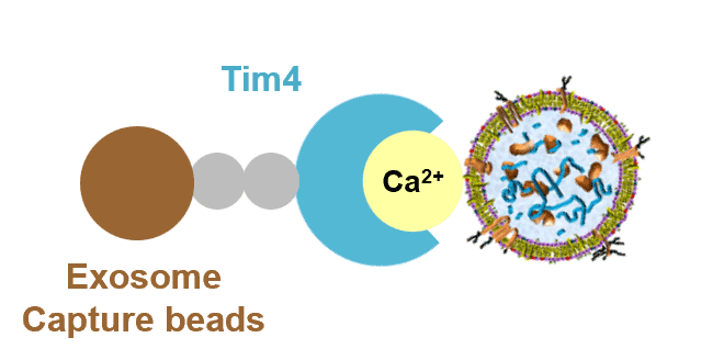
Principle
The PS affinity method, Fujifilm Wako’s proprietary EV isolation technique, utilizes the Tim4 protein, which specifically binds to phosphatidylserine (PS) present on the surface of EVs1). The high specificity of the PS-Tim4 interaction enables to capture high-purity EVs in their intact state.
(1) Isolation of EVs
EVs are isolated from the sample by Exosome Capture Beads.
(2) Staining of EVs
EVs are stained with fluorescent-labeled anti-EV marker antibodies.
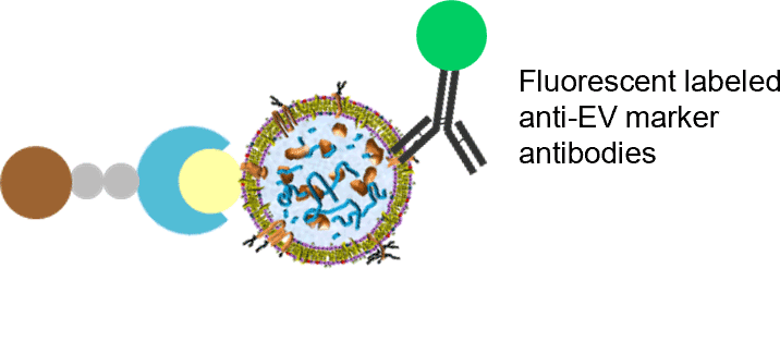
(3) Flow cytometry analysis
Flow cytometry analysis is performed with EVs bound to magnetic beads.
About commercial or for-profit purposes
Please use this product for research purposes only. If you wish to use it for commercial or for-profit purposes, please contact Fujifilm Wako at ffwk-labchem-tec@fujifilm.com.
Protocol (for 2 reactions)
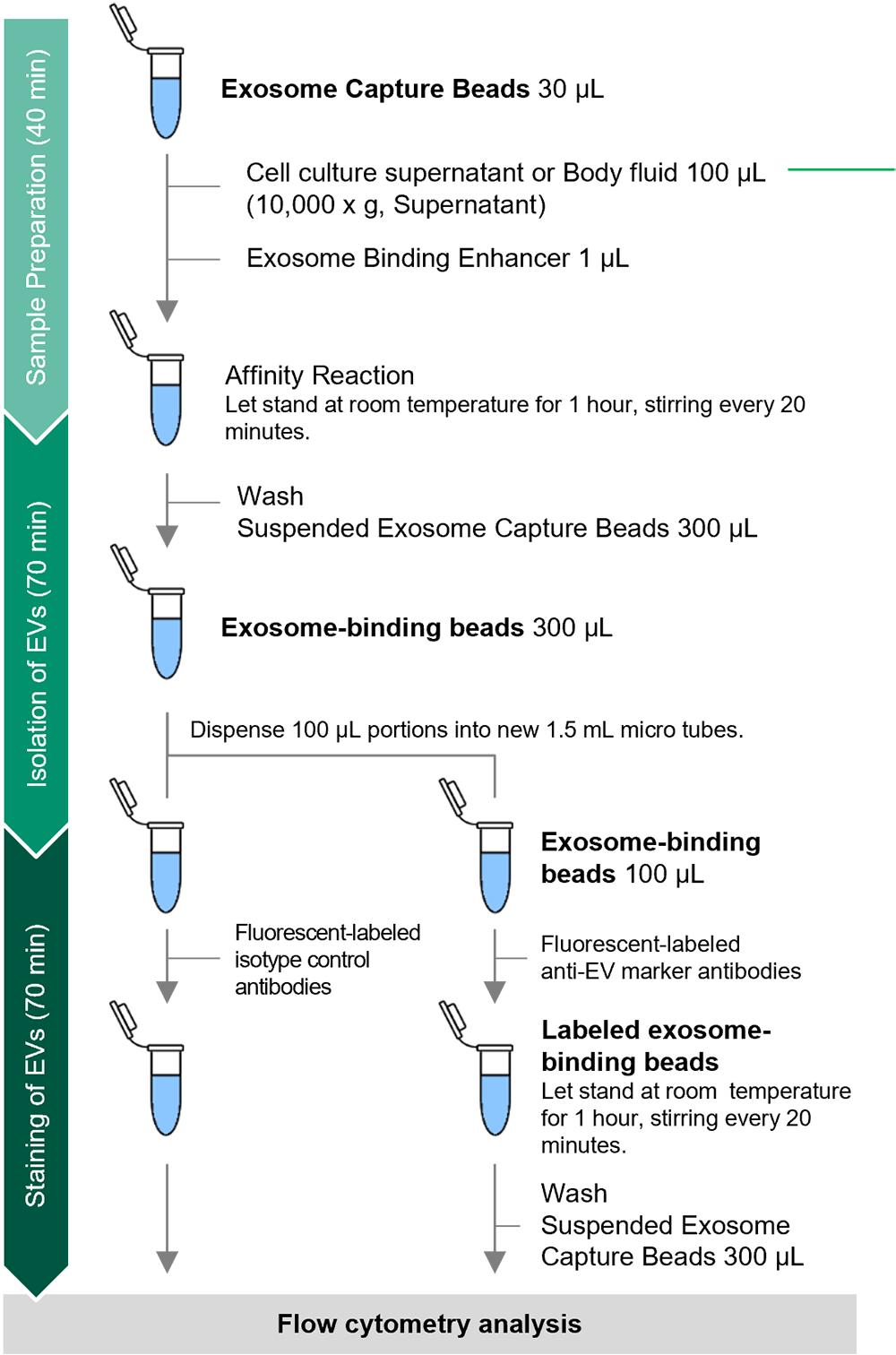
Number of Reactions and Sample Volume
In the EV isolation process, a standard protocol is set for 1.5 mL microtubes (for two reactions). To increase the scale of reactions, the amount of Exosome Capture Beads and sample volume should be scaled up according to the table below.
| Number of Reactions |
Exosome Capture Beads (μL) |
Sample Volume (μL) |
|---|---|---|
| 2 reactions (basic) | 30 | 100 |
| 3 reactions | 40 | 133 |
| 4 reactions | 50 | 167 |
| 5 reactions | 60 | 200 |
| 6 reactions | 70 | 233 |
| 7 reactions | 80 | 267 |
| 8 reactions | 90 | 300 |
| 9 reactions | 100 | 333 |
| 10 reactions | 110 | 367 |
Data
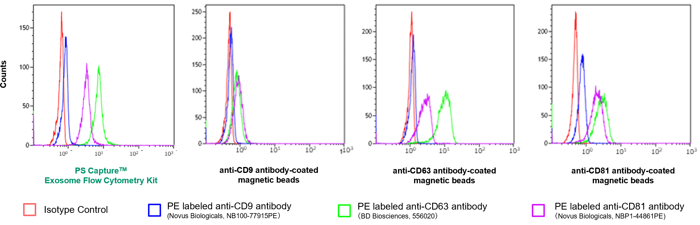
Analysis of Surface Antigens of EVs in Cell Culture Supernatants
EVs in K562 cell culture supernatants were immobilized this kit and with other companies' products (anti-CD9, CD63, and CD81 antibody-coated magnetic beads), and fluorescent-labeled anti-CD9, CD63, and CD81 antibodies were bound for analysis of EV surface antigen by flow cytometry.
Reference Data -Comparison of Signal/Background Ratios-
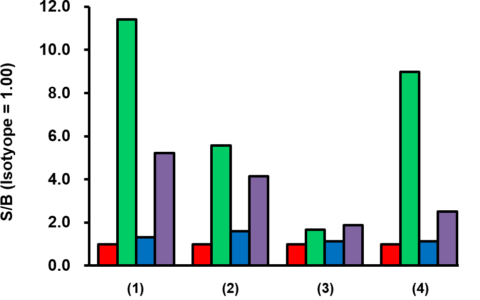
(1) PS Capture™ Exosome Flow Cytometry Kit
(2) Competitor’s product: Anti-CD81 antibody-coated magnetic beads
(3) Competitor’s product: Anti-CD9 antibody-coated magnetic beads
(4) Competitor’s product: Anti-CD63 antibody-coated magnetic beads
[Result]
The PS Capture™ Exosome Flow Cytometry Kit was able to detect EV surface antigens with high sensitivity.
Analysis of Surface Antigens of EV in Serum and Plasma
EVs contained in 100 μL of human serum, heparin plasma, and EDTA plasma (buffer-exchanged) were immobilized onto magnetic beads. Detection of EVs was performed using PE-labeled anti-mouse IgG isotype control and PE-labeled anti-human CD9 antibody.
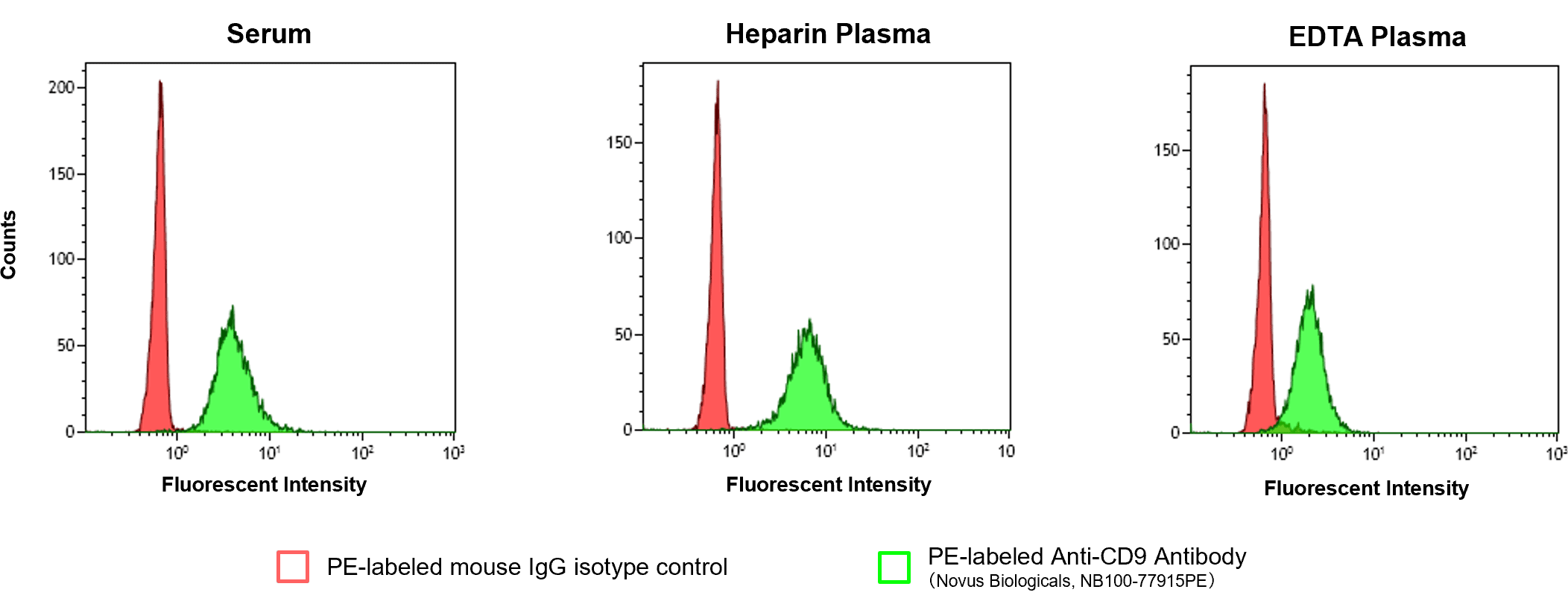
[Result]
Using this product, CD9 in human serum and plasma (heparin/EDTA) was detected.
Detection of proteins inside EVs by Flow Cytometry
EVs purified from HEK293T cell culture supernatant were permeabilized with EV-Perm™ Permeabilization Pretreatment Kit for Exosome Membrane (Product No. 294-85701), and Alix that is the protein inside EVs, and CD63 that is EV surface marker protein were detected using this kit.
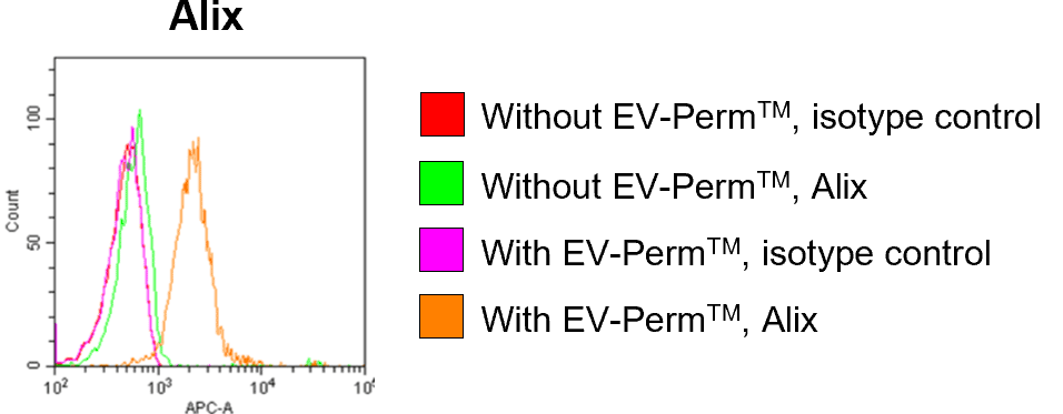

[Result]
With permeabilization, Alix inside EVs was detected by flow cytometry.
References
- Nakai, W. et al.: Sci. Rep., 6(1), 1(2016).
A novel affinity-based method for the isolation of highly purified extracellular vesicles
FAQ
About Sample
- What animal species are suitable for this kit?
- The kit has been used with samples derived from humans, mice, cows, and monkeys.
- What is the minimum amount of samples for detection?
- Approximately 33 µL is required per sample. However, if the amount of EVs in the sample is small, pretreat the cell culture supernatant by centrifugation and concentrate by ultrafiltration to prepare samples (recommended filter: Sartorius Vivaspin 20, molecular weight cut-off 100 K, Product No. VS2041).
- Is there a way to measure purified EV samples?
- Dilute the purified EVs to an appropriate concentration, and proceed to 2, “Isolation of Extracellular Vesicles," in the instruction manual. To dilute purified EVs, use the Washing Buffer (10 x) supplied with the kit diluted 10-fold with ultrapure water. Purified exosomes can be detected at concentrations of 125-1,000 ng/mL.
About Kit Components and Protocol
- Is it possible to use the MagCapture™ Exosome Isolation Kit PS Ver.2 (Product No. 290-84103) for flow cytometry?
- It is not possible. Use the PS Capture™ Exosome Flow Cytometry Kit, in which the magnetic beads and the composition of wash buffer are optimized for flow cytometry.
- The MagCapture™ Exosome Isolation Kit PS Ver.2 includes a bead-Tim4 binding step. Why does this product not include a binding step?
- This product directly binds Tim4 to magnetic beads, whereas the MagCapture™ Exosome Isolation Kit PS Ver.2 binds it via the biotin-avidin system. The specifications differ because each product uses a different type of magnetic bead.
- Are antibodies for EV detection included in this kit?
- Fluorescent-labeled antibodies for EV detection are not included in this kit. Please purchase an appropriate fluorescent-labeled antibody.
- Is it possible to use fluorescent-labeled secondary antibodies for detection?
- It is possible. After isolating EVs, follow the protocol below for primary antibody reaction, secondary antibody reaction, and flow cytometry analysis.
- Mix unlabeled primary antibody and Exosome Capture Beads, and allow to stand at room temperature for 1 hour. Vortex the magnetic beads for about 5 seconds at 20 min, 40 min, and 1 hour.
- Wash twice with 300 µL of WB (+ Enhancer).
- Dilute PE-labeled secondary antibody from the Jackson ImmunoResearch Laboratories (Product No. 115-115-164) 100-fold with WB (+ Enhancer) and add to the magnetic beads in 2.
- Allow to stand at room temperature for 1 hour. Vortex the magnetic beads for about 5 seconds at 20 min, 40 min, and 1 hour.
- Wash 3 times with 300 µL of WB (+ Enhancer).
- Suspend the magnetic beads in 300 µL of WB (+ Enhancer).
- Analyze by flow cytometry.
About Application
- Is quantitative analysis possible with this kit?
- For quantitative analysis, the number of Exosome Capture factors per bead particle needs to be determined. This product is not guaranteed for it, and quantitative analysis is not possible.
Troubleshooting
- Multiple Exosome Capture Beads and EVs are bound, causing the magnetic beads to aggregate.
- Set the gate for singlet bead fraction based on the forward and side-scatter light plots, and detect the fluorescent signal of Exosome Capture Beads within the gate. For a typical sample, the singlet bead fraction represents 50-70% of the total. Additionally, using a cell strainer may reduce bead aggregation.
- The magnetic beads do not stick to the side of the magnetic stand.
- The magnetic beads in this kit are optimized for flow cytometry, and the concentration of the magnetic beads is low. It may be difficult to visually confirm whether the magnetic beads are collected. In the washing step, place the tube on the magnetic stand for at least 1 minute, and then slowly pipet so as not to aspirate the magnetic beads.
Overview / Applications
| Outline | This kit is a reagent that can qualitatively analyze extracellular vesicles contained in cell culture supernatants and body fluid samples by flow cytometry. After extracellular vesicles are reacted and immobilized on magnetic beads on which extracellular vesicle surface phosphatidylserine (PS) specifically binding protein is immobilized, fluorescent labeling for any extracellular vesicle marker protein by using antibodies, we can detect marker proteins on extracellular vesicle surface with high sensitivity. Before using it, prepare the primary antibody for each surface marker protein, the fluorescent dye-labeled secondary antibody, or the primary antibody labeled with fluorescent dye. |
|---|
Property
Manufacturer Information
Alias
For research use or further manufacturing use only. Not for use in diagnostic procedures.
Product content may differ from the actual image due to minor specification changes etc.
If the revision of product standards and packaging standards has been made, there is a case where the actual product specifications and images are different.