Phos-tag™ Fluorescent Gel Stain Reagents
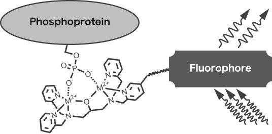
Phos-tag™ fluorescent gel stain reagents are fluorescent stain reagents for SDS-PAGE gels. They applied with functional molecule "Phos-tag™" that captures phosphate groups(-PO32-). By staining the gel after SDS-PAGE with them, phosphorylated proteins can be stained specifically. Because they can be used even at physiological pH, it allows detecting proteins that are phosphorylated by histidine (pHis) and aspartic acid (pAsp), which are susceptible to hydrolysis when acidic.
FUJIFILM Wako offers different wavelengths: Yellow, Magenta, Cyan and Aqua.
Features
- Highly sensitivity
- Staining at physiological pH
- High selectively to pSer, pTyr, pHis, and pAsp residues
- No need for any radioactive material, labeling with a chemical or antibody
- Completed within 2 hours
-
Competitor Product A
- pH:2-4
- 5 steps
(fixing・washing・staining・destaining・washing) - The time required:≧ 5 hours
- Solution exchange:11 times
- The detection limit of ovalbumin:>5 ng/lane
-
Phos-tag™ Fluorescent Gel Stain Reagent
- pH:7-8 (no dephosphorylation occurs)
- 3 steps
(fixing・staining・washing) - The time required:≦ 2 hours
- Solution exchange:4 times
- The detection limit of ovalbumin:>1 ng/lane
How to use
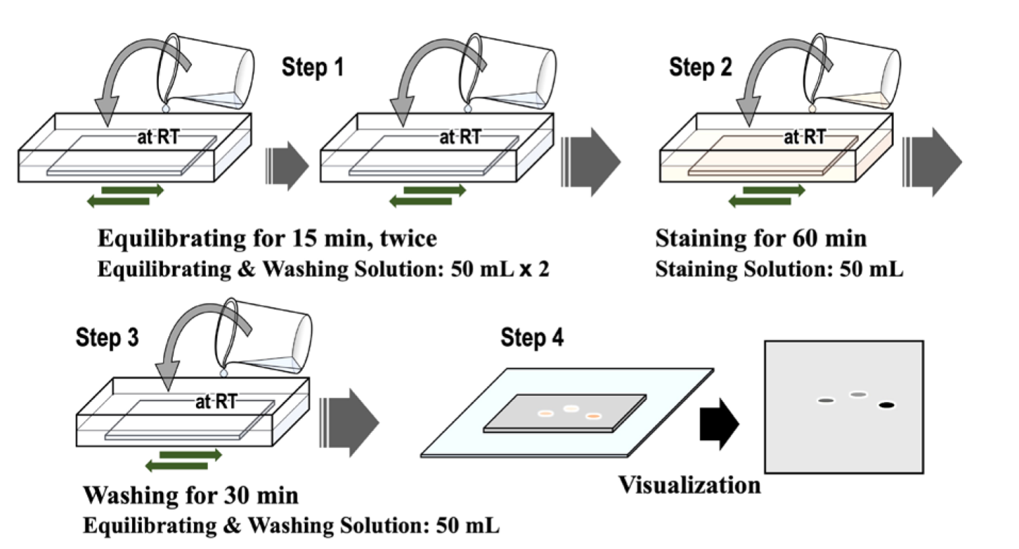
- Configuration of Equiliprating & Washing Solution as well as Staing Solution requires the Mixed reagents for Phos-tag™ Common Solution 5x (product code:383-15231).
References
“Quantitative monitoring of His and Asp phosphorylation in a bacterial signaling system by using Phos-tag Magenta/Cyan fluorescent dyes”, Emiko Kinoshita-Kikuta, Hiroshi Kusamoto, Syogo Ono, Keisuke Akayama, Yoko Eguchi, Masayuki Igarashi, Toshihide Okajima, Ryutaro Utsumi, Eiji Kinoshita, Tohru Koike, Electrophoresis, 2019
Excitation wavelength / fluorescence wavelength
-
Phos-tag™ Yellow
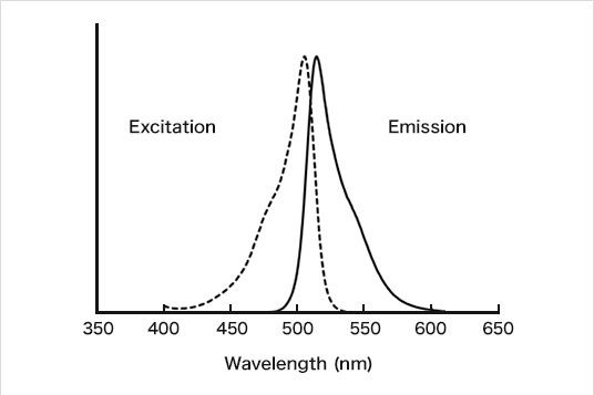
Ex/Em=505nm/514nm
-
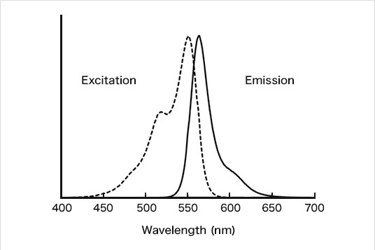
Phos-tag™ Magenta
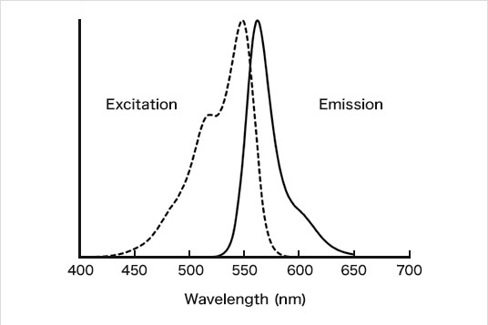
Ex/Em=547nm/561nm
-
Phos-tag™ Cyan
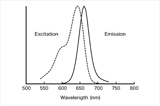
Ex/Em=643nm/661nm
-
Phos-tag™ Aqua
Ex/Em=551nm/564nm
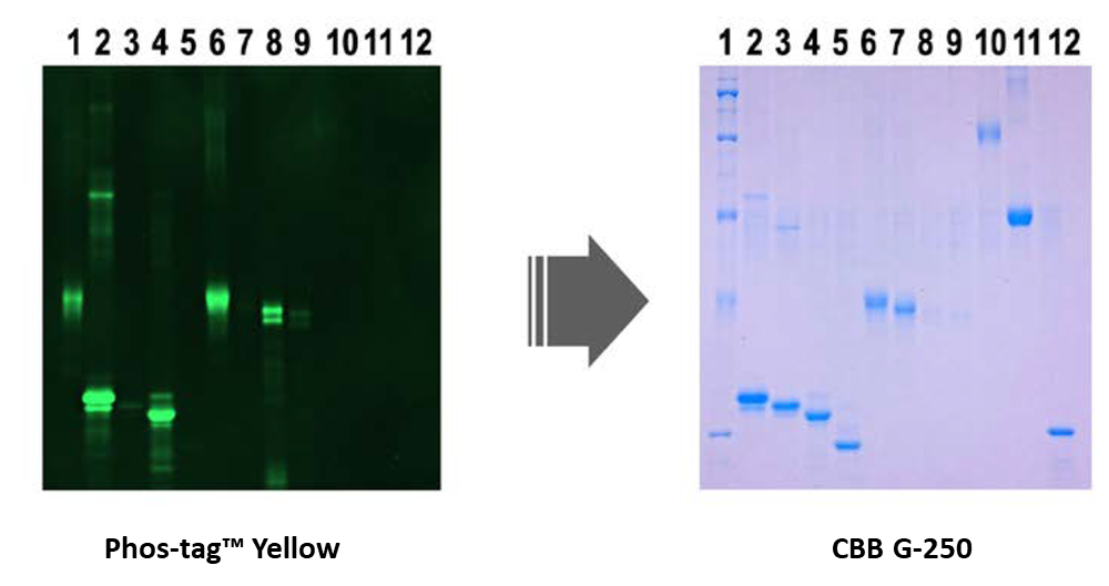
Application Data [1]
Staining phosphorylated and non-phosphorylated proteins
-
-
Lane No. ( ) indicates the number of Phosphate groups
- Marker
- α-casein (9P)
- ALP-treated α-casein
- β-casein (5P)
- ALP-treated β-casein
- Ovalbumin (2P)
- ALP-treated ovalbumin
- Pepsin (1P)
- ALP-treated pepsin
- ß-galactosidase
- Bovine serum albumin
- Carbonic anhydrase
Selectivity of the Phos-tag™ Yellow stain: A 10% (w/v) polyacrylamide gel containing four sets of intact and ALP(alkaline phosphatase)-treated phosphoproteins (1 μg each of α-casein, β-casein, ovalbumin, and pepsin) was stained with Phos-tag™ Yellow (left) and subsequently with CBB G-250 gel satin (right).
Phosphorylated proteins were specifically detected after SDS-PAGE.
Data provided by Emiko Kinoshita-Kikuta, Eiji Kinoshita, Tohru Koike, Department of Functional Molecular Science, Institute of Biomedical and Health Sciences, Hiroshima University
Application Data [2]
Analysis of phosphorylation of histidine and aspartic acid residues by Phos-tag™ fluorescent gel staining
Signal transduction from EnvZ, histidine kinase, to OmpR: The regulator responses play a role in bacterial gene expression. Phosphorylation of histidine and aspartic acid residues were analyzed on autophosphorylated EnvZ and OmpR phosphorylated by EnvZ.
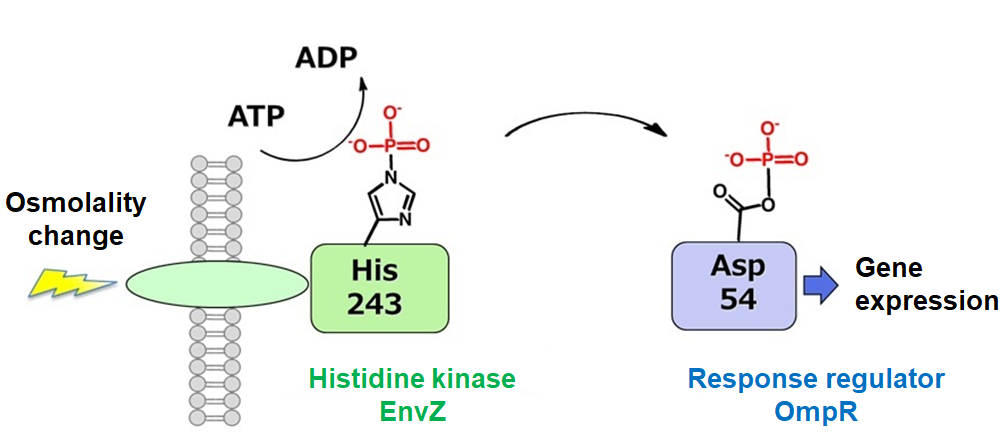
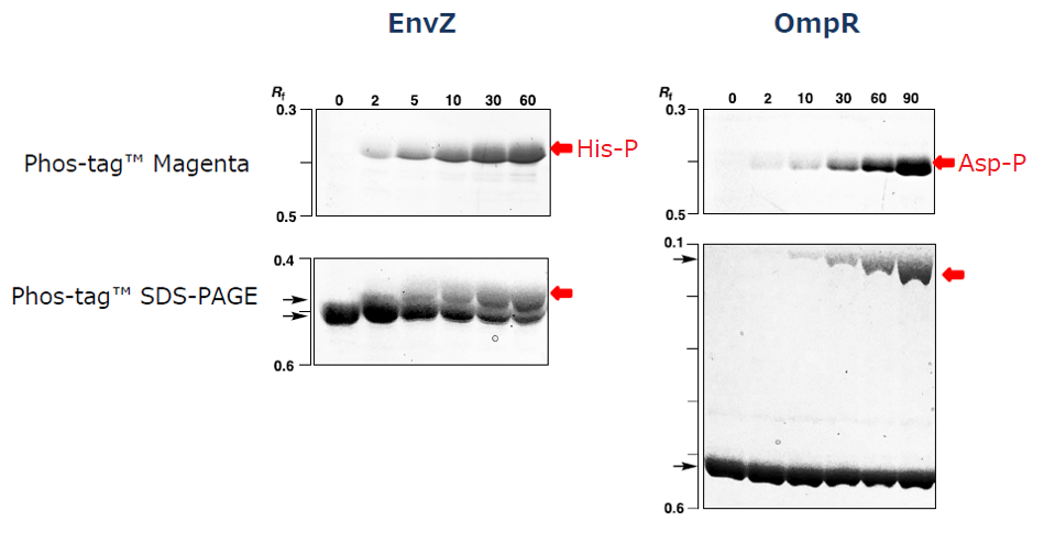
Both Phos-tag™ Magenta and Phos-tag™ SDS-PAGE showed that phosphorylated histidine (His) and aspartic acid (Asp) residues were increased with a prolonged incubation time of the related kinase. Especially, Phos-tag™ Magenta is able to detect exclusively phosphorylated His or Asp residues.
Data provided by Emiko Kinoshita-Kikuta, Eiji Kinoshita, Tohru Koike, Department of Functional Molecular Science, Institute of Biomedical and Health Sciences, Hiroshima University
Application Data [3]
Screening for histidine kinase inhibitors
Screening for histidine kinase inhibitors was performed by using Phos-tag™ fluorescent gel staining.
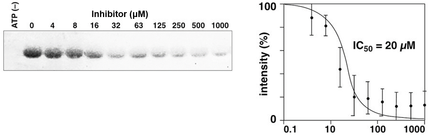
Phosphorylation was suppressed dependent on the histidine kinase inhibitor concentration.
Data provided by Emiko Kinoshita-Kikuta, Eiji Kinoshita, Tohru Koike, Department of Functional Molecular Science, Institute of Biomedical and Health Sciences, Hiroshima University
Product List
- Open All
- Close All
For research use or further manufacturing use only. Not for use in diagnostic procedures.
Product content may differ from the actual image due to minor specification changes etc.
If the revision of product standards and packaging standards has been made, there is a case where the actual product specifications and images are different.