SuperSep™ Ace
SuperSep™ Ace is a polyacrylamide gel for electrophoresis with high resolution and long-term storage stability. It can be used not only in general SDS-PAGE, but also in electrophoresis of DNA. With no gel in SDS, it can also be used in native-PAGE. In addition, SuperSep™ Ace Mini, which is smaller than normal gels and allows more rapid electrophoresis (approximately 20 minutes), is currently available.
Products
| SuperSep™ Ace | SuperSep™ Ace Mini | ||
|---|---|---|---|
| Plate size | 100 x 100 x 3 (mm) | 100 x 60 x 4 (mm) | |
| Gel size | 90 x 85 x 1 (mm) | 82 x 50 x 1 (mm) | |
| Number of wells | 13 | 17 | 17 |
| Well Volume (μL) | 30 | 25 | 20 |
| Recommended electrophoresis chamber | EasySeparator™ (Product Number:292-36411) |
EasySeparator™ Mini (Product Number:051-09251) |
|
Running Buffers
| Product Number | |||
|---|---|---|---|
| Proteins | Sample buffer (×2) | 0.125M Tris-HCl, pH6.8、20% Glycerol、4% SDS、10% 2-Mercaptoethanol、0.004% BPB | 196-11022 |
| Running buffer (×10) | 0.25M Tris、1.92M Glycine、1% SDS | 184-01291 | |
| Low molecular weight proteins | Sample buffer (×2) | 0.125M Tris-HCl, pH6.8、20% Glycerol、4% SDS、10% 2-Mercaptoethanol、0.004% BPB | 196-11022 |
| Running buffer (×10) | 0.5M Tris、0.5M Tricine、1% SDS | 200-17071 | |
| Nucleic acids | Sample buffer (×2) | 0.01M Tris-HCl, pH7.9、1mM EDTA、30% Sucrose、0.004% BPB or Loading Buffer | 313-90111 |
| Running buffer (×10) | 0.25M Tris、1.92M Glycine or TBE buffer | 318-90041 | |
SuperSep™ Ace
Features
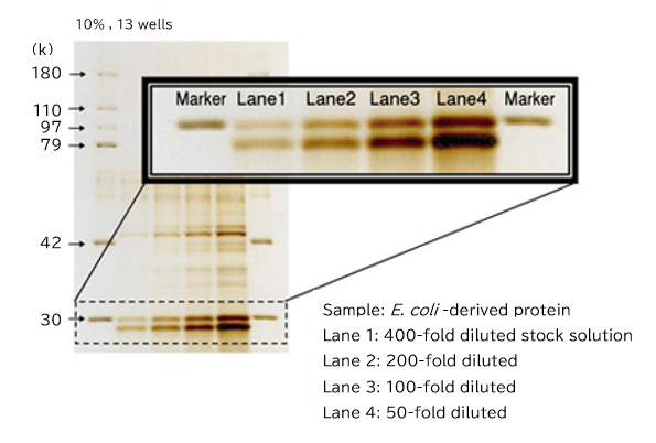
- Good separation and clear bands (below)
- Can be used with multichannel pipette
- Patch on the side of the box to describe the gel type
Recommended electrophoresis conditions
- Constant current: 20 mA/gel
- Constant voltage: 200 V
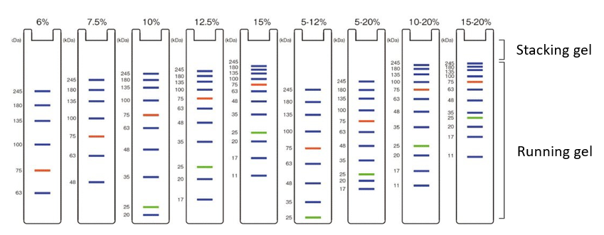
Electrophoretic pattern by concentration
Select the acrylamide concentration based on the electrophoretic patterns presented below.
WIDE-VIEW™ Prestained Protein Size Marker III (Product Number: 230-02461) is used.
Examples of use
Example1 Electrophoresis of gel filtration-purified fractions for E. coli-expressed His-tagged protein
SuperSep™ was used for electrophoresis of gel filtration-purified fractions for E. coli-expressed His-tagged protein (35 kDa) to identify fractions containing the target protein.
Fraction No. 84 to 96 had a band at 35 kDa, showing that E. coli-expressed His-tagged protein was contained in Fractions No. 84 to 96.
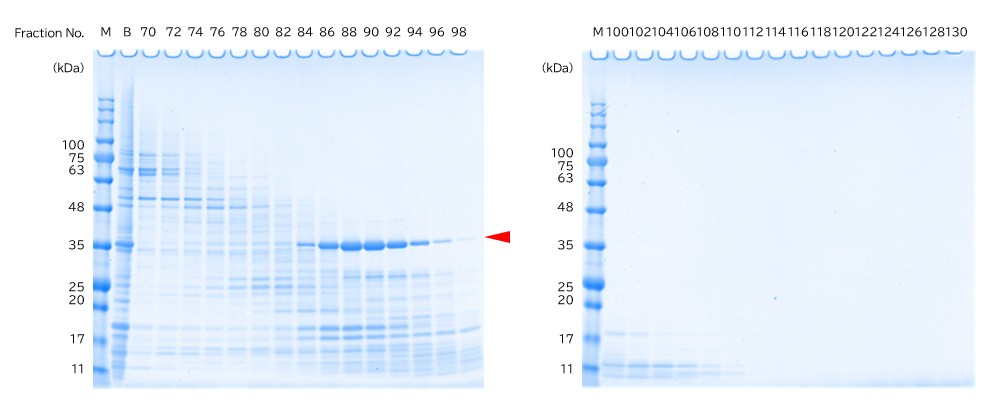
*2 B: Protein before gel filtration
| Gel Running buffer Sample buffer Sample Molecular weight marker Stain |
:SuperSep™ Ace 10-20%, 17 well (Product Number: 198-15041) :SDS-PAGE Buffer, pH 8.5 (Product Number: 192-16801) :Sample Buffer Solution (containing 3-mercapto-1, 2-propanediol) (×4) (Product Number: 196-16142) :Gel filtration fractions for E. coli-expressed His-tagged protein (35 kDa) :WIDE-VIEW™ Prestained Protein Size Marker III (Product Number: 230-02461) :Quick-CBB PLUS (Product Number: 178-00551) |
Example2 Determination of purified protein concentration
SuperSep™ was used for electrophoresis of E. coli-expressed His-tagged protein purified as described above in Example1 and albumin solutions to determine the concentration of purified protein.
The thickness of the bands detected showed that the concentration of purified E. coli-expressed His-tagged protein was approximately 0.45 mg/mL.
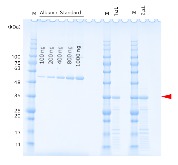
| Gel Running buffer Sample buffer Sample Molecular weight marker Stain |
:SuperSep™ Ace 10-20%, 17 well (Product Number: 198-15041) :SDS-PAGE Buffer, pH 8.5 (Product Number: 192-16801) :Sample Buffer Solution (containing 3-mercapto-1, 2-propanediol) (×4) (Product Number: 196-16142) :E. coli-expressed His-tagged protein (purified protein) and 2 mg/mL albumin solution, bovine serum-derived (Product Number: 015-25613) :WIDE-VIEW™ Prestained Protein Size Marker III (Product Number: 230-02461) :Quick-CBB PLUS (Product Number: 178-00551) |
Examples of use in native-PAGE
- Analysis of the phospholipase C-δ1 pleckstrin homology domain using native polyacrylamide gel electrophoresis
Analytical Biochemistry, Volume 431, Issue 2, 15 December 2012, Page 106–114 - Core–shell clusters of human haemoglobin A and human serum albumin: artificial O2-carriers having various O2-affinities J. Mater. Chem. B, 2015, Advance Article
- Bacterial toxin-inducible gene expression of cathelicidin-B1 in the chicken bursal lymphoma-derived cell line DT40: Functional characterization of cathelicidin-B1 Peptides, Volume 59, September 2014, Pages 94–102
SuperSep™ Ace Mini
Features
- Electrophoresis is completed in approximately 20 minutes (constant voltage: 300 V)
- Useful to confirm protein expression or purification
- High storage stability
Examples of use
SuperSep™ Ace Mini was used for electrophoresis of IgG antibody solutions. It was demonstrated that 20 minutes of electrophoresis resulted in good separation of markers and proteins in individual samples.。
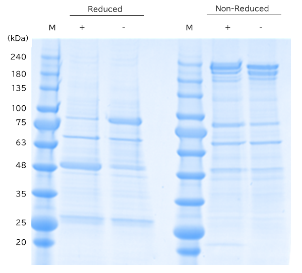
| Gel Running buffer Sample Molecular weight marker Stain Electrophoresis conditions |
:SuperSep™ Ace Mini, 10-20%, 17 well (Product Number: 191-18613) :SDS-PAGE Buffer, pH 8.5 (Product Number: 192-16801) :Reduced/non-reduced IgG antibody solutions (+: heat-treated, -: non heat-treated) :WIDE-VIEW™ Prestained Protein Size Marker III (Product Number: 230-02461) :Quick-CBB PLUS (Product Number: 178-00551) :Constant voltage (320 V), 20 minutes |
Secret technique for time-saving
CBB staining using a microwave
A secret technique for completing the entire process from electrophoresis using SuperSep™ Ace Mini*1 to CBB staining to confirm protein expression in approximately 40 minutes*2 is described below. Please try it.
- 1 SuperSep™ Ace can also be used.
- 2 The time required depends on the experimental conditions.
Reagents and instruments required
- Quick-CBB PLUS (Product Number: 174-00553)
- Microwave
- Plastic wrap
- Kimwipes®
- Heat-resistant tray for holding gel
Procedure
- SDS-PAGE
- Staining using a microwave: 1 minute x 2 to 4 times
Soak the gel in staining solution and then cover it with plastic wrap for 1-minute heating in a microwave (500 W). Repeat this procedure until the entire gel becomes colored. - Decolorization using a microwave: 1 minute x 4 to 6 times
Discard the staining solution in the tray and pour 100 mL of deionized water to soak the gel. Heat the tray with rounded Kimwipes® inside in a microwave (500 W) for approximately 1 minute and then replace deionized water. Repeat the same procedure several times.*3
- 3 The gel under the solution is not broken by bumping. To remove the background, leave the gel in deionized water overnight.
- Note:
- : If the gel is completely covered with plastic wrap, make several holes with a needle. Since the tray becomes very hot, use gloves when removing it.
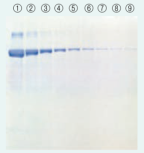
Staining results
-
Conventional method
-
Microwave method
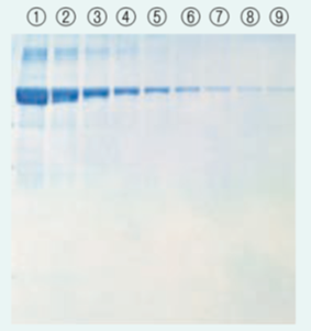
Sample:BSA
Amount of sample:
① 5 μg as total protein
② to ⑨ Two-fold serial dilution
Product List
- Open All
- Close All
SuperSep™ Ace
SuperSep™ Ace Mini
For research use or further manufacturing use only. Not for use in diagnostic procedures.
Product content may differ from the actual image due to minor specification changes etc.
If the revision of product standards and packaging standards has been made, there is a case where the actual product specifications and images are different.