SeeDB2
Drs. Ke and Imai developed a new type of tissue clearing agent, SeeDB2. SeeDB2 is ideal for high-resolution three-dimensional imaging of fluorescent proteins. SeeDB2G and S are designed match the refractive indices of glycerol (1.46) and oil (1.52), minimizing spherical aberrations for high-NA glycerol- and oil- immersion objective lenses, respectively. SeeDB2 is particularly powerful in confocal and super-resolution microscopy using high-NA objective lenses. It is noteworthy that many of fluorescent proteins are highly preserved in SeeDB2, much better than in PBS or other commercialized mounting media optimized for fluorescent dyes. Thus, SeeDB2 is also an ideal mounting medium for samples labeled with fluorescent proteins. SeeDB2 is useful not only for thick brain tissues, but also for thin samples for cell biology or tissue sections.
Data provided by Drs. Meng-Tsen Ke and Dr. Takeshi Imai, RIKEN
Features
- Easy-to-use
- No special equipment required
- Compatible with IF, FP and other fluorescent labels
- High-resolution imaging
- Not quench fluorescent proteins
Application
SeeDB2 Applications
| Fixation | Clearing | Imaging | |||||
| Fixation 4% PFA/PBS(-) |
Step 1. Permeabilization solution |
Step 2. Clearing Solution 1 |
Step 3. Clearing Solution 2 |
Step 4. Clearing Solution 3 (SeeDB2G) |
Step 5. Clearing Solution 4 (SeeDB2S) |
Mounting SeeDB2G or SeeDB2S |
|
| Processing Temperature | 4 °C | RT | RT | RT | RT | RT | RT |
| Processing Time | 1 day | 12 - 16 hrs | 6 - 24 hrs | 6 - 10 hrs | > 12 hrs | > 12 hrs | |
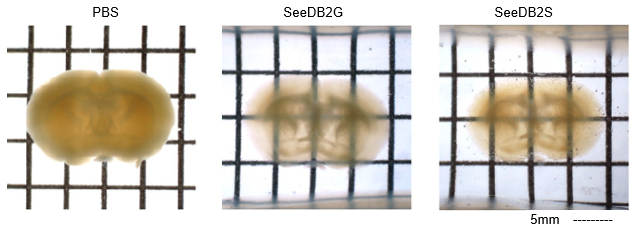
Figure1 . Mouse brain slice (adult, 1.5mm-thick) before and after clearing with SeeDB2.
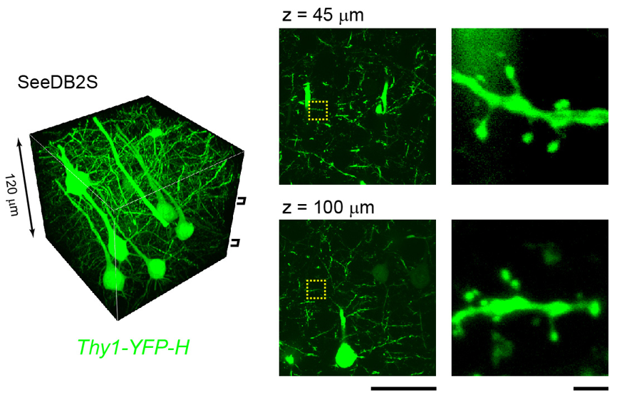
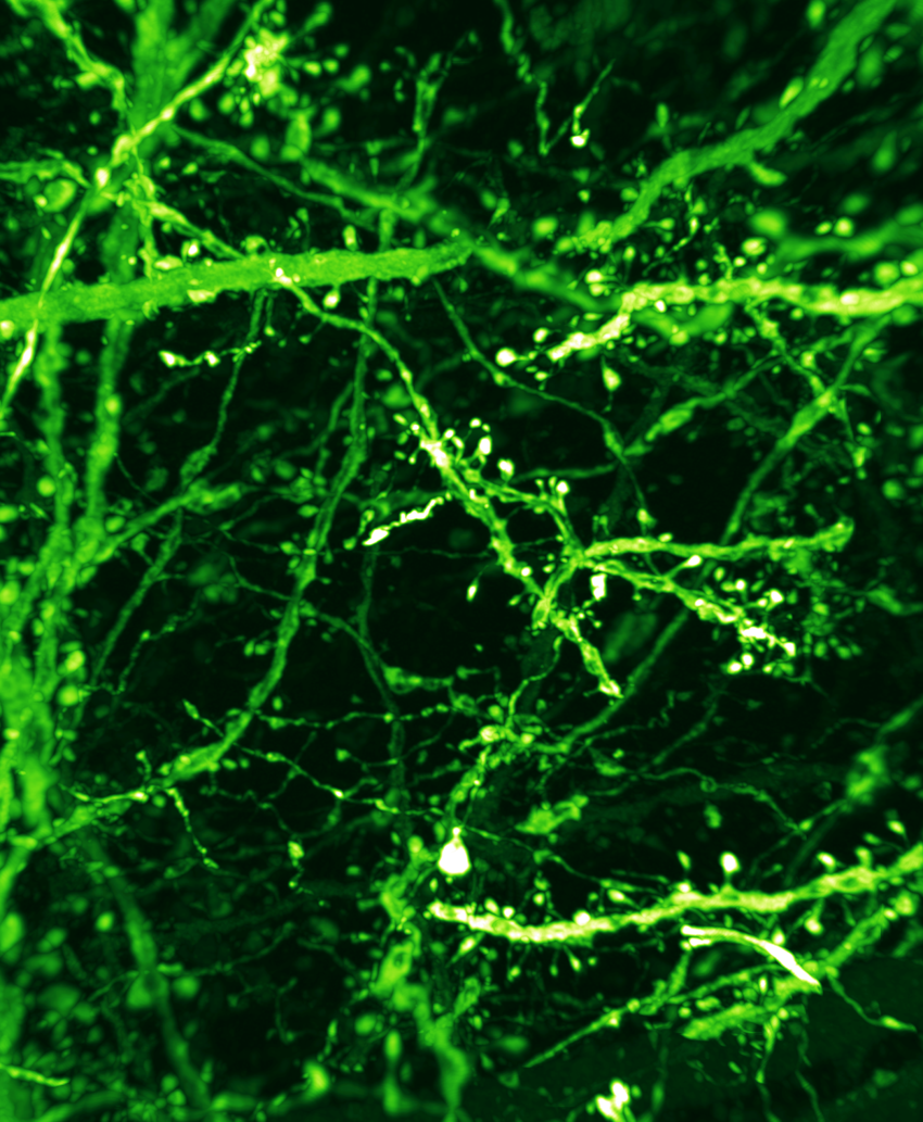
Figure 2. Confocal images of brain slices from a Thy1-YFP (line H) mouse. An oil-immersion objective lens (NA 1.4) was used. Note that fluorescence level did not decrease even though laser power was constant throughout depth. Scale bars are 2μm.
SeeDB
Related Product -SeeDB-
Dr. Takeshi Imai. et al. developed a water-based optical clearing reagent, SeeDB (See Deep Brain), which clears fixed brain samples in a few days without quenching many types of fluorescent dyes, including fluorescent proteins and lipophilic neuronal tracers. SeeDB is a saturated solution of fructose in water with thioglycerol. This method facilitates comprehensive and quantitative analyses for understanding neuronal circuitry, both in the adult and developing mouse brain.
Features of SeeDB
- Easy-to-use
- Plasma membrane and ultrastructure remain intact
- No special equipment required
- Compatible with IF, FP and other fluorescent labels
SeeDB applications
| Fixation | Clearing | Imaging | ||||||
| Fixation 4% PFA/PBS(-) |
Step 1. SeeDB:20w/v% Fructose Solution |
Step 2. SeeDB:40w/v% Fructose Solution |
Step 3. SeeDB:60w/v% Fructose Solution |
Step 4. SeeDB:80w/v% Fructose Solution |
Step 5. SeeDB:100w/v% Fructose Solution |
Step 6. SeeDB | Mounting SeeDB2 |
|
| Processing Temperature | 4 °C | RT | RT | RT | RT | RT | RT | RT |
| Processing Time | 1 day | 4 - 8 hrs | 4 - 8 hrs | 4 - 8 hrs | > 12 hrs | > 12 hrs | 24 hrs | |
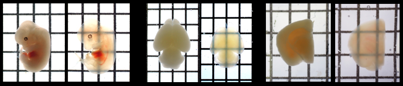
Figure.1 Mouse embryo and mouse brain before and after treatment with SeeDB
 |
||
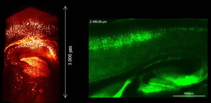
| Figure 2. Imaging of Thy1-YFP(H Line) mouse brain after SeeDB process using Multiphoton microscope and Multiphoton dedicated objective :OLYMPUS, model:XLPLN10XSVMP. | Figure 3. Imaging of Thy1-YFP(H Line) mouse brain after SeeDB process using Confocal microscope and objective :OLYMPUS, UPLSAPO 10X2. | |
See Through Chamber
Related Product - See Through Chamber -
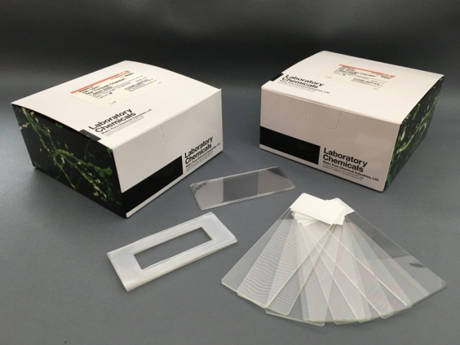
The See through chamber is an observation container used when observing tissue cleared samples with a microscope. The See through chamber is composed of 10 sets of silicon rubber sheets, cover glasses, slide glasses processed to make it easy to observe tissue cleared samples. There are five kinds of thickness of silicone rubber which becomes the spacer, 0.3 mm, 0.5 mm, 1.0 mm, 2.0 mm, and 3.0 mml, and it is possible to select the optimum size according to your samples. The silicone rubber sheet has a protective sheet affixed on both sides, it can be used in close contact with slide glass and cover glass by peeling off at the time of use.
References
- Ke, M. T., Fujimoto, S. and Imai, T. : Nat Neurosci ,6 (8),1154 (2013).
- Ke, M. T., Fujimoto, S. and Imai, T.: Bio-protocol 4(3), e1042 (2014).
- Ke, M. T., and Imai, T. : Curr Protoc Neurosci ,66,2.22.1-2.22.19 (2014).
- Ke et al.,: Cell Reports 14, 2718(2016)
Product List
- Open All
- Close All
For research use or further manufacturing use only. Not for use in diagnostic procedures.
Product content may differ from the actual image due to minor specification changes etc.
If the revision of product standards and packaging standards has been made, there is a case where the actual product specifications and images are different.