EV-Save™ Extracellular Vesicle Blocking Reagent
Extracellular vesicles (EVs), including exosomes, are prone to decrease in quantity during experimental procedures for several reasons. Especially, adsorption to tubes and other surfaces is a significant factor in the loss of EVs. The MISEV2023 guidelines also mention that purified EVs are prone to adsorption to storage containers1).
Using low protein-binding tubes can be effective in preventing the adsorption of EVs to tubes2-3). However, the plastic products used in experiments are not limited to tubes, but also include pipette tips, centrifugal concentrator tubes, etc., and it is impractical to pretreat all these consumables for low adsorption.
In such situations, a blocking agent added to samples or purified EV solutions offers an excellent solution in terms of operability and versatility. The EV-Save™ Extracellular Vesicle Blocking Reagent (Product Number: 058-09261), which contains a polymer, can be added to samples or purified EV solutions to prevent their adsorption to tubes, pipette tips, and centrifugal concentrator tubes. Furthermore, EV-Save™ acts as a cryoprotectant, minimizing damage to EVs during freeze-thaw cycles. Fujifilm Wako has also developed EV-Save™ which is specialized for in vivo experiment and EV purification by Tangential flow filtration (TFF).
What is EV-Save™?
EV-Save™ Extracellular Vesicle Blocking Reagent is a blocking reagent that suppresses the adsorption of extracellular vesicles (EVs), such as exosomes, to microtubes, pipette tips, and TFF (tandem flow filtration) filter membranes. Simply adding it to samples or purified EV solutions will suppress adsorption. It also acts as a cryoprotectant, minimizing damage to EVs during freeze-thaw cycles. Fujifilm Wako has a lineup of EV-Save™ products specialized for each application.
Product Lineup
Suppress EV adsorption
to tubes, pipette tips, etc.
+
Protect EVs
from freeze-thaw damage
Suppress EV adsorption
and administered to animals
+
Protect EVs
from freeze-thaw damage
suppress EV adsorption
on TFF filtration membranes
+
Protect EVs
from freeze-thaw damage
EV-Save™ Extracellular Vesicle Blocking Reagent
EV-Save™ Extracellular Vesicle Blocking Reagent is a polymeric reagent that prevents EV from adsorbing to tubes, pipette tips, and other labware. It prevents loss of EVs due to adsorption during processing and storage, thereby increasing the yield. Addition of this product is highly recommended prior to ultrafiltration, purification, and storage.
Features
- Prevents adsorption of EV in cell culture supernatants and purified EV onto laboratory equipment
- Protects EV from freeze-thaw damage
- Easy to operate, just add to samples
Sample (Recommended)
- Cell culture supernatant
- Purified EV
Impacts on Analysis
No interference with the analysis of EVs in the following analyses:
- Nanoparticle Tracking Analysis (NTA)
- Western blotting
- ELISA
- Microarray
- Cell culture
Data
Anti-adsorption Effect
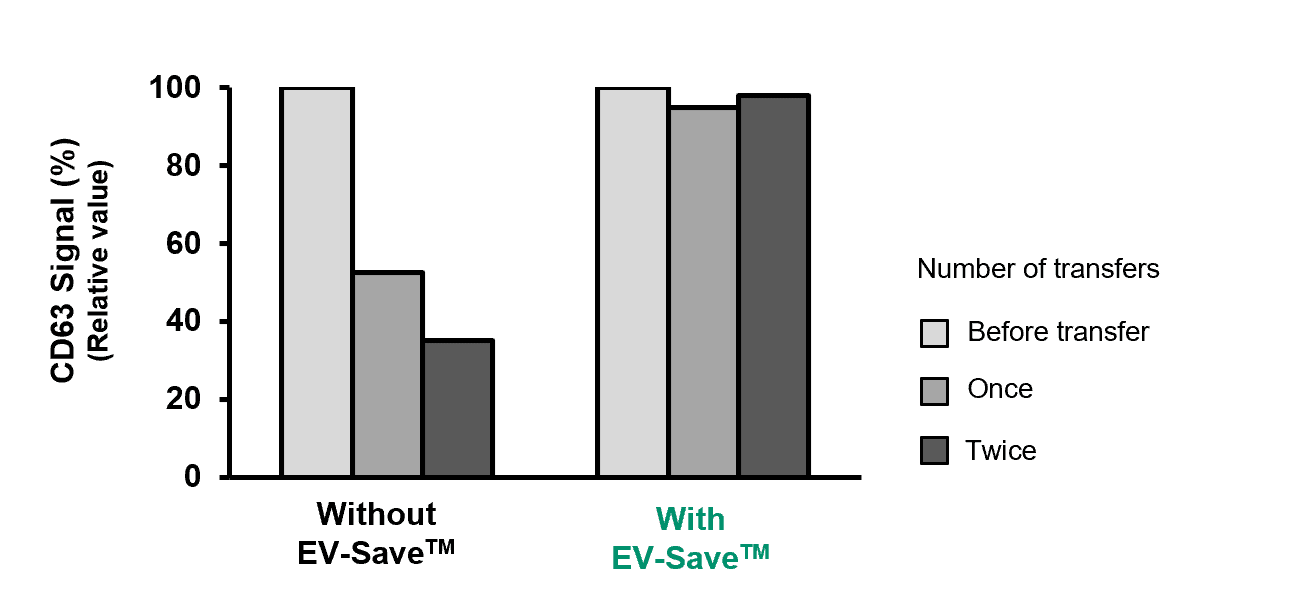
EV-Save™ was added to COLO201 cell-derived EVs purified by the PS affinity method and allowed to stand for 3 minutes. The sample was then transferred to another tube, and the transfer was repeated. The amounts of EV (CD63 signal) in the sample tubes were measured by PS Capture™ Exosome ELISA Kit (Anti Mouse IgG POD).
[Result]
CD63 signal decreased with transfer. The addition of EV-Save™ almost completely prevented the decrease in signal caused by the transfer.
Cryoprotection Effect
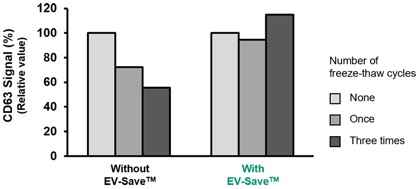
EV-Save™ was added to COLO201 cell-derived EVs purified by the PS affinity method and freeze-thawed. The amounts of EV (CD63 signal) in the sample tubes were measured by PS Capture™ Exosome ELISA Kit (Anti Mouse IgG POD), and their structure was analyzed by transmission electron microscopy (TEM).
(1) ELISA
(2) Transmission electron microscopy
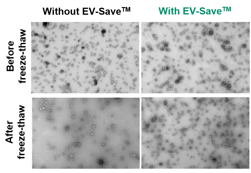
[Result]
The CD63 signal decreased with freeze-thawing. The addition of EV-Save™ prevented the decrease in signal caused by freeze-thawing. Observation by electron microscopy confirmed that the reduction in particle numbers was suppressed.
EV-Save™ Extracellular Vesicle Blocking Reagent for in vivo
EV-Save™ Extracellular Vesicle Blocking Reagent for in vivo applications contains only components that have been used as pharmaceutical additives. This EV anti-adsorption and cryoprotectant can be used for administration of EVs to laboratory animals.
Features
- Prevents adsorption of EV in cell culture supernatants and purified EV onto laboratory equipment
- Protects EV from freeze-thaw damage
- Easy to operate, just add to samples
- Contains only ingredients that have been used as pharmaceutical additives
Sample (Recommended)
- Cell culture supernatant
- Purified EV
Impacts on Analysis
No interference with the analysis of EVs in the following analyses:
- Nanoparticle Tracking Analysis (NTA)
- Western blotting
- ELISA
- Cell culture
Data
Anti-adsorption Effect
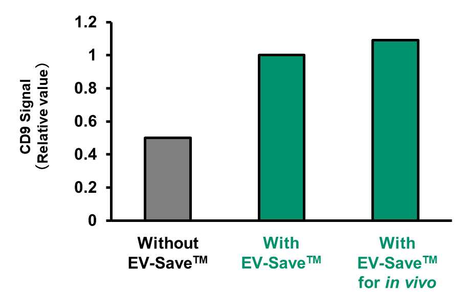
EV-Save™ and EV-Save™ for in vivo applications were added to COLO201 cell-derived EVs purified by the PS affinity method and stored at 4°C for 16 hours. The amounts of EV (CD9 signal) in the sample tubes were measured by PS Capture™ Exosome ELISA Kit (Streptavidin HRP).
[Result]
CD9 signal decreased without the addition of EV-Save™. The addition of EV-Save™ or EV-Save™ for in vivo applications prevented the decrease in signal.
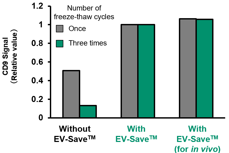
Cryoprotection Effect
EV-Save™ or EV-Save™ for in vivo applications was added to COLO201 cell-derived EVs purified by the PS affinity method and freeze-thawed once or three times. The amounts of EV (CD9 signal) in the sample tubes were measured by PS Capture™ Exosome ELISA Kit (Streptavidin HRP).
[Result]
In the absence of EV-Save™, freeze-thawing reduced the CD9 signal. This signal reduction was prevented by the addition of EV-Save™ or EV-Save™ for in vivo applications.
EV-Save™ Extracellular Vesicle Blocking Reagent for TFF
Tangential flow filtration (TFF) is widely used as an effective method for concentrating extracellular vesicles (EVs) and performing buffer exchange. By flowing the sample parallel to the filtration membrane, TFF minimizes sample loss by preventing accumulation on the membrane surface. However, for samples such as EVs, which are prone to nonspecific adsorption, recovery may be reduced due to adsorption to the membrane and physical stress during filtration.
EV-Save™ Extracellular Vesicle Blocking Reagent for TFF is a specialized reagent developed to improve EV recovery by mitigating physical stress and suppressing membrane adsorption during TFF processing. Use of this product enables efficient and consistent recovery of EVs.
Features
- Suppresses EV damage and adsorption to the filtration membrane during EV purification, concentration, and buffer exchange using TFF
- Simple protocol: add directly to the sample and equilibration buffer
- Prevents EV loss due to adsorption to tubes and protects against freeze-thaw damage, enabling direct storage of EV solutions after TFF. (Note: This effect is achieved only when this product is added during buffer exchange.)
Sample (Recommended)
- Cell culture supernatant
- Purified EV
Data
Improved Recovery during Buffer Exchange of Purified EVs
EVs were purified from the culture supernatants of bone marrow-derived mesenchymal stem cells (BMMSC) and adipose-derived mesenchymal stem cells (ADMSC) using the MassivEV™ EV Purification Column PS. Buffer exchange was then performed using a Repligen TFF system with HBS buffer containing this product at a 1:100 dilution. EV recovery after buffer exchange was evaluated using the PS Capture™ Exosome ELISA Kit (Streptavidin-HRP), with anti-CD63 or anti-CD81 antibodies used as detection antibodies. Particle size distribution was assessed using nanoparticle tracking analysis (NTA).
(1) ELISA
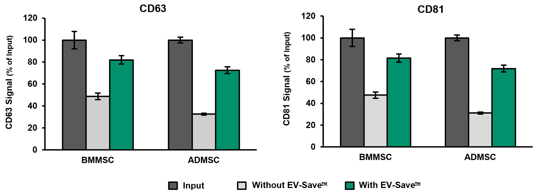
[Result]
The addition of this product improved EV recovery during TFF-based buffer exchange.
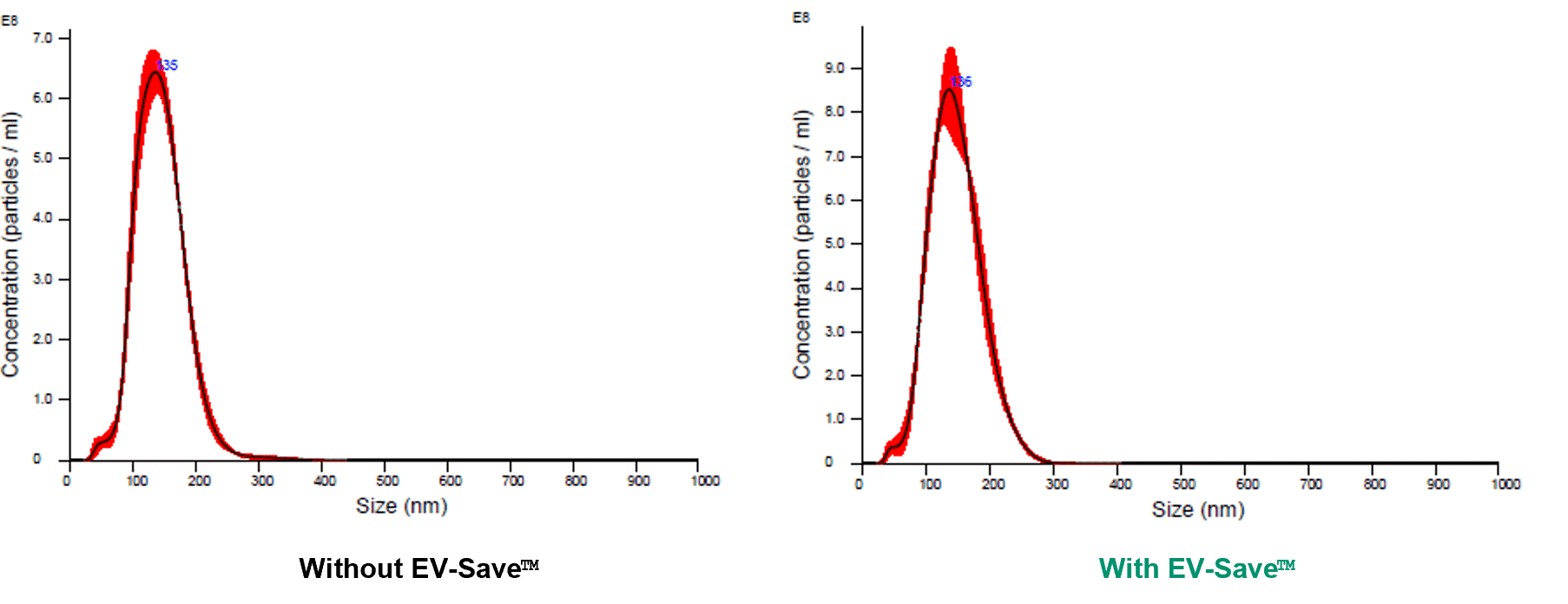
(2) NTA
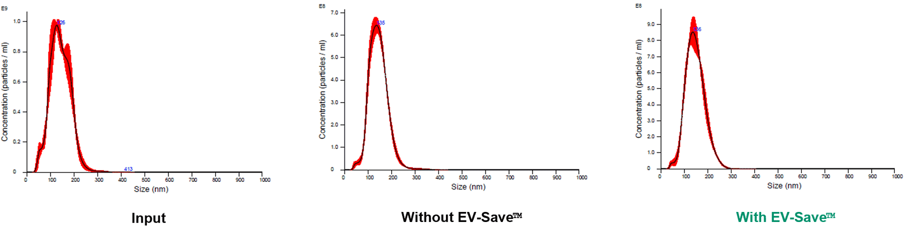
[Result]
No significant change in EV particle size distribution was observed by the addition of this product.
Improved Recovery during TFF-Based EV Purification from Cell Culture Supernatants
Cell culture supernatants were first concentrated 20-fold using TFF, followed by a 100-fold buffer exchange. This product was added at a 1:100 dilution to both the cell culture supernatant and the replacement buffer. EV recovery was measured using ELISA with anti-CD63 and anti-CD81 antibodies. Particle size distribution was analyzed using NTA.
(1) ELISA
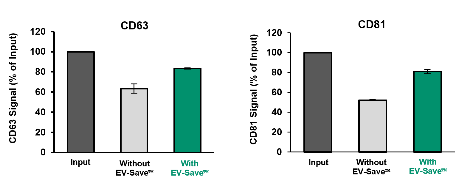
[Result]
The addition of this product improved EV recovery during EV purification from cell culture supernatants.
(2) NTA
[Result]
No significant change in EV particle size distribution was observed by the addition of this product.
References
- Welsh, J. A. et al.: J. Extracell. Vesicles, 13(2), e12404(2024).
Minimal information for studies of extracellular vesicles (MISEV2023): From basic to advanced approaches - Evtushenko, E. G. et al.: PloS One, 15(12), e0243738(2020).
Adsorption of extracellular vesicles onto the tube walls during storage in solution - 「決定版エクソソーム実験ガイド」, ed. by Yoshioka, Y. and Ochiya, T., Yodosha, Japan, (2004). (Japanese)
Product List
- Open All
- Close All
Standard type
For in vivo
For TFF
For research use or further manufacturing use only. Not for use in diagnostic procedures.
Product content may differ from the actual image due to minor specification changes etc.
If the revision of product standards and packaging standards has been made, there is a case where the actual product specifications and images are different.
The prices are list prices in Japan.Please contact your local distributor for your retail price in your region.