Blocking Reagents (Exosome)


Exosomes are known to adsorb to the wall of plastic tubes, which considerably affects the exosome yield and other outcomes. In addition, as exosomes are damaged by freeze and thaw, protective agents suitable for storing exosomes have been desired. With this background in mind, Fujifilm Wako developed the EV-Save™ series of adsorption prevention/cryoprotection reagents for exosomes. EV-Save™ considerably inhibits adsorption of exosomes to the tube walls and protects exosomes from the freeze-thaw damage.
Product Line-up
More Information
Exosome Loss: Causes and Troubleshooting
Extracellular vesicles (EVs), including exosomes, are prone to decrease in quantity during experimental procedures for several reasons. Commonly identified causes include: (1) degradation of the EVs themselves, (2) breakdown or denaturation of marker proteins used for detection, (3) aggregation or fusion of EVs, and (4) adsorption of EVs to tubes and other surfaces. Among these, adsorption to tubes and other surfaces is a significant factor in the loss of EVs. The MISEV2018 guidelines also mention that purified EVs are prone to adsorption to storage containers1).
A study by Evtushenko et al. demonstrated that HT-29 cell-derived EVs isolated using ultracentrifugation and stored in PBS at 4°C showed a decrease in concentration over time, with a loss of about 50% after 48 hours2), highlighting the significance of this effect.
In our own research at Fujifilm Wako, we have also observed a decrease in the signal of CD63, a marker protein of EVs, with repeated transfers to new tubes. Even after a single transfer, the CD63 signal decreased by about 50% (Figure 1).
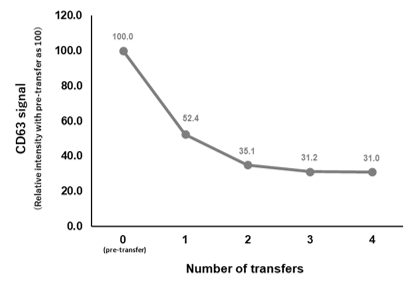
Figure 1 Decrease in EV marker proteins (CD63) due to tube transfer
EVs derived from COLO201 cells were purified using the PS affinity method and then added to a tube. After standing, the EVs were transferred to a new tube. The transfer was repeated four times. Subsequently, the CD63 signal was measured using the PS Capture™ Exosome ELISA Kit (Anti-Mouse IgG POD) (Product Number: 297-79201).
Using low protein-binding tubes can be effective in preventing the adsorption of EVs to tubes2-3). However, the plastic products used in experiments are not limited to tubes, but also include pipette tips, centrifugal concentrator tubes, etc., and it is impractical to pretreat all these consumables for low adsorption. Indeed, at Fujifilm Wako, we have confirmed the loss of EVs even when using centrifugal concentrator tubes for ultrafiltration. In the abovementioned study by Evtushenko et al., blocking the tubes with bovine serum albumin (BSA) or EVs themselves was effective2). However, the authors also note that this approach cannot be highly recommended due to the labor involved in preparation and the potential impact on subsequent analysis.
In such situations, a blocking agent added to samples or purified EV solutions offers an excellent solution in terms of operability and versatility. Fujifilm Wako’s EV-Save™ Extracellular Vesicle Blocking Reagent (Product Number: 058-09261, hereinafter referred to as EV-Save™), which contains a polymer, can be added to samples or purified EV solutions to prevent their adsorption to tubes, pipette tips, and centrifugal concentrator tubes (Figure 2). We have confirmed that the reagent has negligible effect on Nanoparticle Tracking Analysis (NTA), ELISA, Western blotting, microarray, and live cells*1. Additionally, in anticipation of administering EVs for in vivo experiment, Fujifilm Wako has developed the EV-Save™ Extracellular Vesicle Blocking Reagent for in vivo (Product Number 050-09461). This formulation utilizes ingredients with a well-established history as pharmaceutical additives, enabling the selection of the most appropriate reagent based on the intended application*2.
*2 The EV-Save™ Extracellular Vesicle Blocking Reagent for in vivo is not suitable for use with ultrafiltration using centrifugal concentrator tubes.
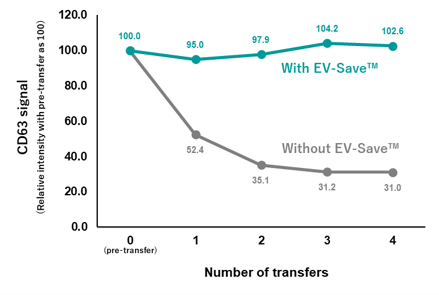
Figure 2 Prevention of Adsorption of EVs by EV-Save™
EVs derived from COLO201 cells were purified using the PS affinity method and then added to a tube. After standing, the EVs were transferred to a new tube. The transfer was repeated four times. Under the condition “With EV-Save™,” 1/100 volume of EV-Save™ was added to the EV solution. Subsequently, the CD63 signal was measured using the PS Capture™ Exosome ELISA Kit (Anti-Mouse IgG POD) (Product No. 297-79201).
Furthermore, EV-Save™ acts as a cryoprotectant, minimizing damage to EVs during freeze-thaw cycles (Figure 3). Repeated freeze-thaw cycles not only damage EVs but also lead to their loss and alterations in their properties. To avoid such repeated cycles, it is recommended to store EVs in small aliquots at -80°C. Additionally, using EV-Save™ helps to prevent the loss of EVs caused by freeze-thaw cycles.
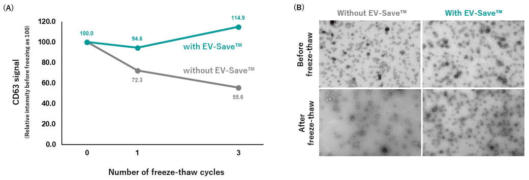
Figure 3 Cryoprotection of EVs by EV-Save™
EVs derived from COLO201 cells were freeze-thawed using the PS affinity method. After being subjected to freeze-thaw cycles, the CD63 signal of these EVs was measured using the PS Capture™ Exosome ELISA Kit (Anti-Mouse IgG POD) (Product No. 297-79201) (A), and their structure was analyzed by transmission electron microscopy (TEM) (B).
The loss of EVs leads to decreased efficiency in experiments and significantly impacts the results of analyses. A thorough understanding of EV characteristics, combined with the implementation of targeted measures, ensures the acquisition of more accurate data.
References
- Théry, C. et al.: J. Extracell. Vesicles, 7(1), 1535750(2018).
Minimal information for studies of extracellular vesicles 2018 (MISEV2018): a position statement of the International Society for Extracellular Vesicles and update of the MISEV2014 guidelines - Evtushenko, E. G. et al.: PloS One, 15(12), e0243738(2020).
Adsorption of extracellular vesicles onto the tube walls during storage in solution - 「決定版エクソソーム実験ガイド」ed. by Yoshioka, Y. and Ochiya, T., Yodosha, Japan, (2004). (Japanese)
For research use or further manufacturing use only. Not for use in diagnostic procedures.
Product content may differ from the actual image due to minor specification changes etc.
If the revision of product standards and packaging standards has been made, there is a case where the actual product specifications and images are different.
The prices are list prices in Japan.Please contact your local distributor for your retail price in your region.