



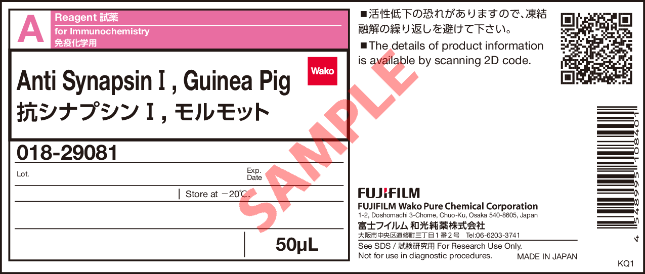
Anti SynapsinⅠ, Guinea Pig
- for Immunochemistry
- Manufacturer :
- FUJIFILM Wako Pure Chemical Corporation
- Storage Condition :
- Keep at -20 degrees C.
-
 Close
Close
- Structural Formula
- Label
- Packing
- SDS
|
Comparison
|
Product Number
|
Package Size
|
Price
|
Availability
|
Certificate of Analysis
|
Purchase |
|---|---|---|---|---|---|---|
|
|
|
50uL
|
|
In stock in Japan |
※Check availability in the US with the distributor.
Document
Product Overview
Synapsin is a synaptic vesicle-associated protein which is thought to regulate synaptic vesicle dynamics. Due to their predominant localization at nerve terminals, Synapsin I are widely used as presynaptic markers in immunohistochemistry. The “Anti Synapsin I, Guinea Pig” is a guinea pig polyclonal antibody, raised against Synapsin I. It can be used to perform multiplex immunohistochemistry1-2).
Antibody Information
| Clonality | Polyclonal |
|---|---|
| Antigen | Rat Synapsin I |
| Host | Guinea pig |
| Formulation | Antiserum |
| Conjugate | Unconjugated |
| Cross-reactivity | Mouse, Rat |
| Application | Immunocytochemistry 1:1,000 Immunohistochemistry (Frozen Section) 1:1,500 |
Protocol (Example)
- Perfusion fixation
- Perfuse with sodium citrate/PBS to remove blood, followed by perfusion with 4 % PFA.
- Post-fixation
- 4 % PFA (-24 hours).
- Immersion in sucrose
- 30 % Sucrose/PBS (4 ℃, from overnight to approximately 2 days)
- Preparation of frozen sections
- After removing sucrose, prepare frozen blocks and cut 30 μm thick sections using a cryostat.
- Glycine treatment
- 25 mM glycine/PBS (Approximately 20 minutes)
- Blocking
- 5 % normal goat serum in 0.3 % Triton X-100/PBS (4 ℃, Overnight)
- Primary antibody reaction
- Anti Synapsin I, Guinea pig (1:1,500), 1 % normal goat serum in 0.3 % Triton X-100/PBS (4 ℃, from overnight to 2 days).
- Washing
- 0.3 % TritonX-100/PBS (5 minutes×3)
- Secondary antibody reaction
- Alexa Fluor® 488 AffiniPure Goat Anti-Guinea Pig IgG (H+L) (1:500), 1 % normal goat serum in 0.3 % Triton X-100/PBS (RT, 2 hours)
- Washing
- 0.3 % TritonX-100/PBS (5 minutes×3)
- Mounting
- The sample is sealed in mounting medium and stored at 4 ℃ in a dark place.
Data
Application Data
Immunohistochemistry
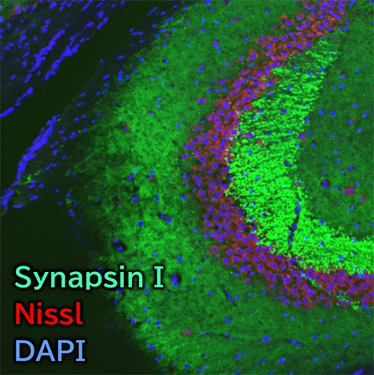
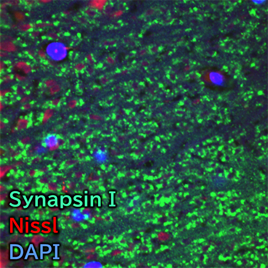
Species: Mouse
Site: Hippocampus (CA3)
Sample: Frozen section
Antibody concentration: 1:1,500
Data by courtesy of
Dr. Miyata, Department of Applied Biology, Kyoto Institute of Technology
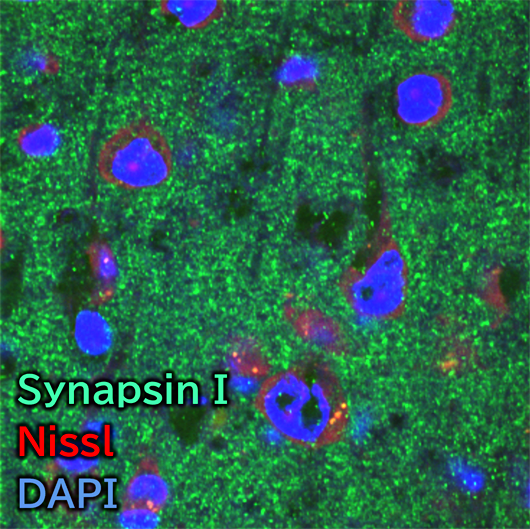
Species: Mouse
Site: Cerebral cortex (Layer V)
Sample: Frozen section
Antibody concentration: 1:1,500
Data by courtesy of
Dr. Miyata, Department of Applied Biology, Kyoto Institute of Technology
Immunocytochemistry
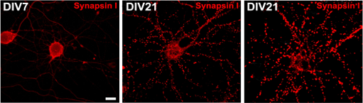
Sample: Rat hippocampus primary cultured neurons
Antibody concentration: 1:1,000
Data by courtesy of
Dr. Miyata, Department of Applied Biology, Kyoto Institute of Technology
[Result]
On day 7 of culture, only a few synapses were observed, whereas by day 21, numerous well-defined punctate synapses were evident. The cultured neurons are metabolically active, and the weak fluorescence observed in the cell bodies likely corresponds to synapsin I either being synthesized in the endoplasmic reticulum or transported along axons.
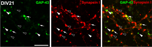
Sample: Rat hippocampus primary cultured neurons
Antibody concentration: 1:1,000
Note: white arrows head → GAP43 positive synapse
black arrows head → Synapsin I positive synapse
Data by courtesy of
Dr. Miyata, Department of Applied Biology, Kyoto Institute of Technology
[Result]
Colocalization of GAP-43 and Synapsin I was observed at synapses (white arrows) in primary rat hippocampal neurons.
References
- Morita, S. and Miyata, S.: Cell Biochem. Funct., 31(5), 400(2013).
Synaptic localization of growth‐associated protein 43 in cultured hippocampal neurons during synaptogenesis - Kawai, S. et al.: Cell Biochem. Funct., 38(4), 392(2020).
Transient increase of microglial C1q expression in the circumventricular organs of adult mouse during LPS‐induced inflammation
FAQ
About protocol
- What secondary antibodies can be used?
- At Fujifilm Wako, the following secondary antibodies have been successfully used:
Alexa Fluor® 488 AffiniPure Goat Anti-Guinea Pig IgG (H+L) (Jackson ImmunoResearch, Product Number: 106-545-003)
About application
- What animals can this antibody be used for?
- Fujifilm Wako has confirmed that it can be used for mice and rats. Other species have not been tested.
Overview / Applications
| Outline | Synapsin are known as phosphorylated proteins specific to neuron. In mammals, three different synapsin genes encode more than eight neuronal isoforms. Synapsin1 is one of the most specific markers of synapses in the central and peripheral nervous systems, and it has been reported that it is present in some sensory nerve endings in addition to synaptic nerve endings. [Antigen] Rat synapsin1 [Species cross reactivity] Mouse and rat [Application] Immunocytochemistry 1:1,000 Immunohistochemistry(frozen section) 1:1,500 |
|---|---|
| Precautions for Use | Avoid repeated freeze and thaw. |
Property
| Appearance | Liquid |
|---|---|
| Composition | Antiserum |
Manufacturer Information
Alias
- SynapsinⅠ antibody
For research use or further manufacturing use only. Not for use in diagnostic procedures.
Product content may differ from the actual image due to minor specification changes etc.
If the revision of product standards and packaging standards has been made, there is a case where the actual product specifications and images are different.