



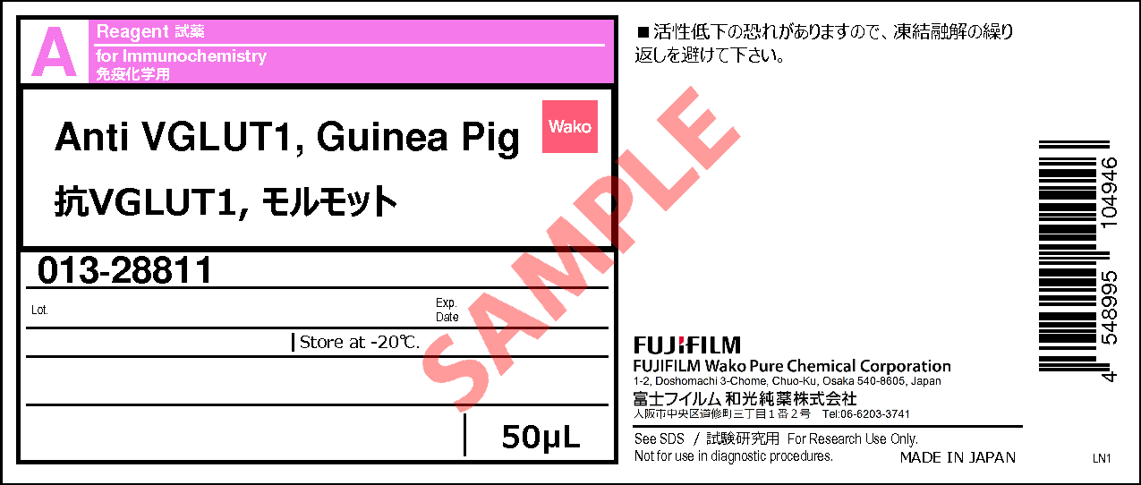
Anti VGLUT1, Guinea Pig
- for Immunochemistry
- Manufacturer :
- FUJIFILM Wako Pure Chemical Corporation
- Storage Condition :
- Keep at -20 degrees C.
-
 Close
Close
- Structural Formula
- Label
- Packing
- SDS
|
Comparison
|
Product Number
|
Package Size
|
Price
|
Availability
|
Certificate of Analysis
|
Purchase |
|---|---|---|---|---|---|---|
|
|
|
50uL
|
|
In stock in Japan |
※Check availability in the US with the distributor.
Document
Product Overview
The vesicular glutamate transporter (VGLUT) mediates the uptake of glutamate—the principal neurotransmitter in mammalian excitatory synaptic transmission—into synaptic vesicles. This process is thought to be driven by the proton electrochemical gradient. In mammals, three isoforms have been identified to date: VGLUT1, VGLUT2, and VGLUT3. These isoforms exhibit distinct expression patterns: VGLUT1 is mainly expressed in the cerebral cortex and hippocampus, whereas VGLUT2 is expressed in the thalamus, hypothalamus, and amygdala. Neurons expressing VGLUT3 are relatively rare, but mRNA expression studies have reported scattered presence in the striatum, hippocampus, cerebral cortex, and raphe nuclei1). In neuroscience research, anti-VGLUT antibodies are widely used as markers for glutamatergic neurons. The “Anti VGLUT1, Guinea Pig” is a guinea pig polyclonal antibody, raised against VGLUT1. It can be used to perform multiplex immunohistochemistry.
Antibody Information
| Clonality | Polyclonal |
|---|---|
| Antigen | Recombinant rat VGLUT1 |
| Host | Guinea pig |
| Formulation | Antiserum diluted in PBS |
| Conjugate | Unconjugated |
| Cross-reactivity | Mouse, Rat |
| Application | Immunohistochemistry (Frozen Section) 1:1,600 |
Protocol (Example)
| 1. Perfusion fixation | Perfuse with sodium citrate/PBS to remove blood, followed by perfusion with 4 % PFA. |
|---|---|
| 2. Post-fixation | 4 % PFA (-24 hours) |
| 3. Immersion in sucrose | 30% Sucrose/PBS (4℃, from overnight to approximately 2 days) |
| 4. Preparation of frozen sections | After removing sucrose, prepare frozen blocks and cut 30 μm thick sections using a cryostat. |
| 5. Glycine treatment | 25 mM glycine/PBS (Approximately 20 minutes) |
| 6. Blocking | 5 % normal goat serum in 0.3 % Triton X-100/PBS (4 ℃, Overnight) |
| 7. Primary antibody reaction | Anti VGLUT1, Guinea pig (1:1,600), 1 % normal goat serum in 0.3% Triton X-100/PBS (4 ℃, from overnight to 2 days). |
| 8. Washing | 0.3 % TritonX-100/PBS (5 minutes x 3) |
| 9. Secondary antibody reaction | Alexa Fluor® 488 AffiniPure Goat Anti-Guinea Pig IgG (H+L) (1:500), 1 % normal goat serum in 0.3 % Triton X-100/PBS (RT, 2 hours) |
| 10. Washing | 0.3 % TritonX-100/PBS (5 minutes x 3) |
| 11. Mounting | The sample is sealed in mounting medium and stored at 4 ℃ in a dark place. |
Data
Application Data
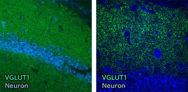
Immunohistochemistry
- Species:
- Mouse
- Site:
- Hippocampus
- Sample:
- Frozen section
- Antibody concentration:
- 1:1,600
[Note]
Neurons were stained with NeuroTrace™ 435/455 Blue Fluorescent Nissl Stain (Thermo Fisher Scientific, Inc.).
Data by courtesy of
Dr. Miyata, Department of Applied Biology, Kyoto Institute of Technology
References
- Gras, C. et al.: J. Neurosci., 22(13), 5442(2002).
A third vesicular glutamate transporter expressed by cholinergic and serotoninergic neurons
FAQ
About protocol
- What secondary antibodies can be used?
- At Fujifilm Wako, the following secondary antibodies have been successfully used:
Alexa Fluor® 488 AffiniPure Goat Anti-Guinea Pig IgG (H+L) (Jackson ImmunoResearch, Product Number: 106-545-003)
About application
- What animals can this antibody be used for?
- Fujifilm Wako has confirmed that it can be used for mice and rats. Other species have not been tested.
Overview / Applications
Property
| Appearance | Liquid |
|---|
Manufacturer Information
Alias
- Anti VGLUT1, Guinea Pig Polyclonal Antibody
For research use or further manufacturing use only. Not for use in diagnostic procedures.
Product content may differ from the actual image due to minor specification changes etc.
If the revision of product standards and packaging standards has been made, there is a case where the actual product specifications and images are different.