



Exosome ELISA Kits


Application of the PS affinity method to ELISA
We developed PS Capture™ Exosome ELISA Kit by applying affinity binding of Tim4 protein with exosomes. This kit is capable of detecting exosomes at a sensitivity higher than that of conventional ELISA methods immobilization of antibodies against exosome surface markers. Exosomes in samples such as culture supernatants and serum are captured by Tim4 protein on adry plate in the presence of calcium ion. The captured exosomes are detected by a primary antibody against an exosome surface marker protein and a labeled secondary antibody. While a mouse anti-CD63 monoclonal antibody is provided with the kit, a user-provided mouse primary antibody against any other exosome surface marker may also be used for exosome detection.
The greatest feature of this kit is that it provides exosomes detection with higher sensitivity than that of Western Blot analysis and conventional product for exosome ELISA. First, the detection limit for exosomes in Western Blotting was examined for comparison with this kit (Figures 1a and b). Western Blot analysis of exosomes purified from COLO201 cells (of human colon adenocarcinoma origin) with an anti-CD63 monoclonal antibody detected exosomes in an amount as small as 75 ng on the protein basis. Next, the detection limits of this kit for exosomes purified from K562 cells (of human leukemia origin) and COLO201 cells were determined to be 49.9 pg and 10.9 pg, respectively, demonstrating that this kit had a detection sensitivity more than 1,000 times higher than that of Western Blotting (Figure 1c). Considering that the detection limits of conventional products for exosome ELISA range approximately several ng to several μg (refer to instruction manuals for individual products), the present results demonstrated that this kit utilizing affinity binding of exosomes to Tim4 via PS has a sensitivity more than 100 times higher than those of conventional ELISA methods involving immobilization of an antibody against an exosome surface protein marker.
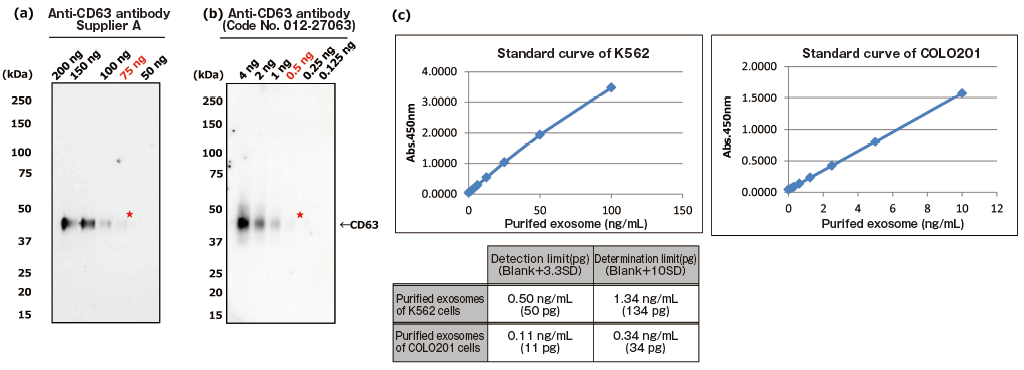
PS ELISA detected the marker proteins with 50-1,000 times higher sensitivity than WB.
| Fig. 1 Comparing the detection sensitivities of Western Blot and PS ELISA | ||
| (a), (b) Result of sensitivity by Western Blot with each of anti-CD63 antibody (supplier A and Wako: Code No. 012-27063). Sample: purified extracellular vesicles from cell culture supernatant of COLO201 cells with MagCapture™ Exosome Isolation Kit PS (Code No. 293-77601) |
||
| ★:detection limit by Western Blot | ||
| (c) Result of limit by PS ELISA | ||
| A standard curve was prepared using blank value of buffer and absorbance value of 2-fold serial dilution samples of extracellular vesicles purified from cell culture supernatant of K562 cells and that of COLO201 cells with MagCapture™ Exosome Isolation Kit PS. Then, the detection limit of purified extracellular vesicles of K562 cells and COLO201 cells were calculated using its standard curve. (each dilution point: n=6, blank: n=12) | ||


PS Capture™ Exosome ELISA Kit (Streptavidin HRP)
-
Features
- Sensitive qualitative analysis (50 to 1000 folds more sensitive than WB in detection)
- Direct relative quantitation of extracellular vesicles in conditioned medium and body fluid specimens
- Application to detection systems using antibodies from various animal species and lectins
- Microanalysis with small amounts of extracellular vesicles
(only 1/10 to 1/1000 of the sample amount for WB)
-
Assay Principle
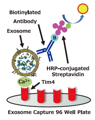
Exosome Capture 96 Well Plate which is pre-coated with protein that specifically binds to phosphatidylserine (PS) on the surface of extracellullar vesicles (EVs) capture EVs with Ca2+, Then biotinylated antibody for the surface marker protein of EVs is used as a primary detection and HRP-conjugated streptavidin is used as a secondary detection.
Application Data
Dilution Linearity of Cell Culture Supernatant and Blood Samples
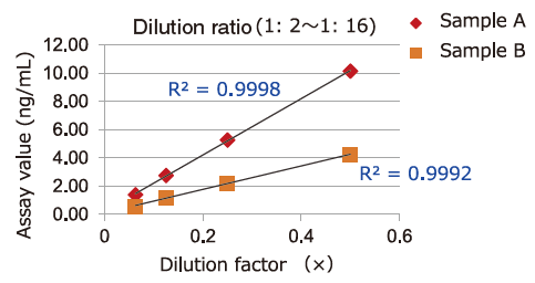
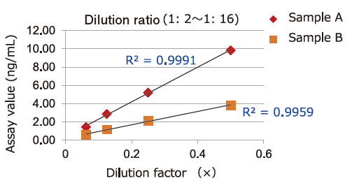
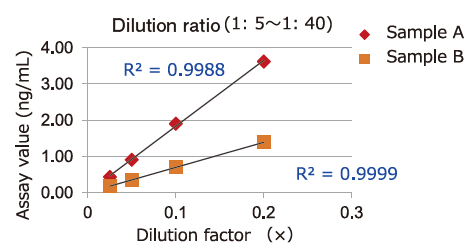
Dilution linearity of four specimens, ① human serum diluent ② human heparin plasma diluent ③ human EDTA plasma diluent (2 specimens, 4-step serial dilution each) and ④ COLO201 cells cell culture supernatant diluent (4-step serial dilution), was assessed by measuring the concentration of exosomes (CD63 detection) with standard curve obtained using extracellular vesicles purified from cell culture supernatant of COLO201 cells.
-
①Serum
-
②Heparin plasma
-
③EDTA plasma
-
④COLO201 cell culture supernatant
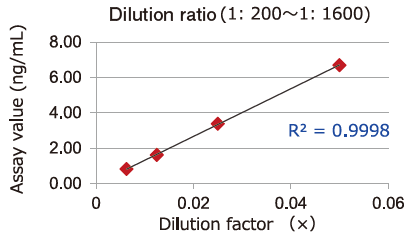
Samples in serum and heparinized plasma have given favorable linearity when diluted 2 folds or more.
Even samples in EDTA-treated plasma will give favorable linearity when diluted 5 folds or more.
Comparison of Capture Ability between PS Affinity and Antibody
Microplate wells, pre-coated with anti-CD antibodies (CD9, CD63, CD81) and PS Affinity (Tim4), were incubated with pretreated ※ 1 cell culture supernatants of iPSC, COLO201 and HEK293T cells. Captured exosomes on the plate were detected using biotinylated anti-CD antibodies (CD9, CD63, CD81).
※ 1 Pretreatment condition: 10,000 x g, 30min.
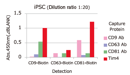
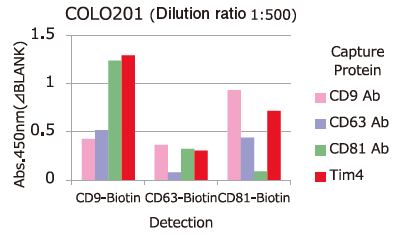
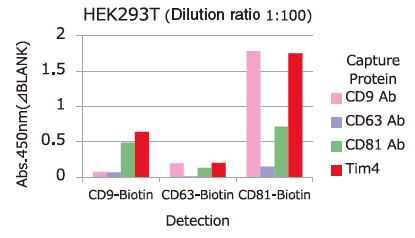
Almost all of the exosomes derived from various cell lines were efficiently captured by Tim4 compared with anti-CD9, 63, 81 antibodies.
Spike and Recovery Assay with Blood Samples【EpCAM】
Pretreated※2 pooled normal human serum, EDTA-treated plasma, and heparinized plasma were spiked with exosomes isolated and purified from COLO201 cell conditioned medium using MagCapture™ Exosome Isolation Kit PS (Code No. 293-77601) at various concentrations. The spiked exosomes in a sample were detected using biotin-labeled anti-EpCAM※3 antibody (MBL), and the recovery rate of exosomes from the sample was calculated based on the standard curve prepared using exosomes purified from COLO201 cell conditioned medium.
※ 2 Pretreatment condition: 10,000 x g, 30min. ※ 3 EpCAM: Epithelial cell adhesion molecule
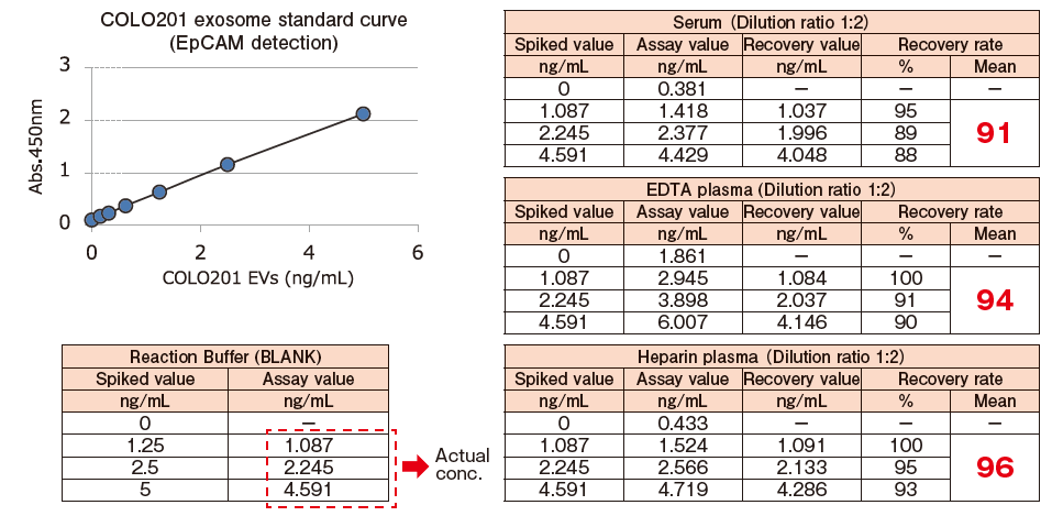
The spiked recovery rates based on the detection of EpCAM fell within a range of 100%±10%, showing favorable recovery performance.
Test of Residual Extracellular Vesicles in EV-depleted FBS
Extracellular vesicles in untreated FBS, commercial EV-depleted FBS and ultracentrifugation (UC)-treated FBS ※ 4 were detected using Biotinylated anti-CD9 antibody ※ 5.
Capture: Tim4 / Detection: Biotinylated anti-CD9 antibody
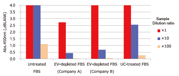
※ 4 Ultracentrifugation condition: 160,000 x g, 16h
※ 5 Anti-CD9 antibody (Code No. 014-27763) was biotinylated using Biotin Labeling Kit-SH (Code No. 348-90941).
PS Affinity ELISA System enables test of residual extracellular vesicles in EV-depleted FBS.
Sugar Chain Analysis by Sandwich ELISA with rBC2LCN Lectin and Tim4
Exosomes in five-fold dilution of pretreated※6 cell culture supernatants of iPS cells were detected using Biotinylated anti-CD63 antibody (Code No. 019-27713) and Biotinylated rBC2LCN lectin※7.
Capture: Tim4 / Detection: Biotin labelled compounds
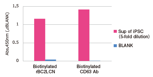
※ 6 Pretreatment condition: 10,000 x g, 30min.
※ 7 rBC2LCN binds specifically to Fucα1-2Galβ 1-3GalNAc (GlcNAc) and is known as a undifferentiated marker of iPS and ES cells. rBC2LCN (Code No. 029-18061) was biotinylated using EZ-Link™ Sulfo-NHS-LC-LC-Biotin (ThermoFisher).
Reference: S. Saito, et al., Sci. Rep., 8, 3997 (2018)
PS Affinity ELISA is applicable for sugar chain analysis on exosomes with a various combination of lectins.
Comparison of recovery efficiency with Polymer Precipitation
From 1 mL of pretreated ※ 8 COLO201 cell conditioned medium, exosomes were isolated and purified by PS affinity method or polymer precipitation method (competiter A) and then measured for signals of CD63, an exosome marker, using PS Capture™ Exosome ELISA Kit (streptavidin HRP). From absorbance of each sample, the amount of residual exosomes in a post-purification sample and the recovery rate of exosomes were determined for comparison.
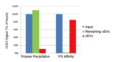
Calculation of recovery efficiency
Input: Measured value of 100-fold diluted conditioned medium※Ⅰ
Remaining sEVs: Measured value of post-purification 100-fold diluted conditioned medium ※Ⅰ
Recovered sEVs: Measured value of 1000-fold diluted purified sample ※Ⅱ
※Ⅰ Measured value of 100-fold diluted sample has been confirmed to fall within the measurement range. In the preliminary investigation, a dilution ratio appropriate for ELISA is recommended to be calculated for each sample
※Ⅱ Exosome samples purified with this kit and polymer reagents are theoretically concentrated 10 folds. (e.g. 1 mL of input → 100μL of purified exosomes) Value obtained by multiplying the concentration ratio by the dilution ratio for ELISA corresponds to integrated dilution ratio of the purified exosome for ELISA
The ELISA system allowing highly sensitive detection of exosom es will ease efficiency comparison among exosome recovery methods and validation of the method.
CD9/63/81-Capture Human Exosome ELISA Kit (Streptavidin HRP)
Features
- Specific to human extracellular vesicles.
- Direct analysis from cell culture sup. or blood samples. No need for isolation.
- Qualitative and quantitative analysis.
Note: Purified extracellular vesicle standard is required for quantification. - Simple operation and high reproducibility
Application Data
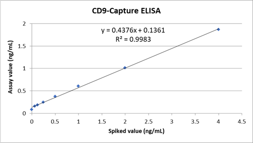
Spike and recovery assay of human derived-extracellular vesicles from mouse EDTA plasma.
Evaluate the assay linearity using mouse EDTA plasma (2-fold diluted) spiked with Exosome, from COLO201 cells, Purified [code No. 052-09301].
-
1. CD9-Capture Human Exosome ELISA Kit
-
2. CD63-Capture Human Exosome ELISA Kit
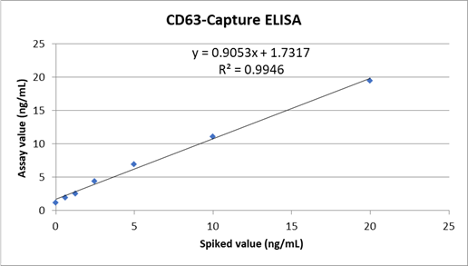
-
3. CD81-Capture Human Exosome ELISA Kit
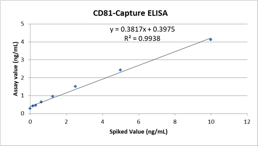
Assay Sample 2-fold diluted mouse EDTA plasma spiked with exosomes purified from COLO201 cells at each concentration. Assay Kit CD9/63/81-Capture Human Exosome ELISA Kit
Human-derived exosomes were measured with good linearity from mouse EDTA plasma sample.
Comparison of extracellular vesicles expression in various human cell lines culture supernatant.
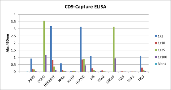
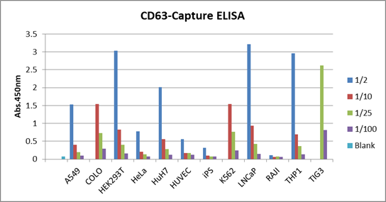
Culture supernatants of 12 types of human-derived cell lines were diluted 2, 10, 25, and 100 fold and measured with this kit.
1. CD9-Capture Human Exosome ELISA Kit
2. CD63-Capture Human Exosome ELISA Kit
3. CD81-Capture Human Exosome ELISA Kit
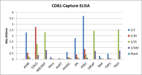
It was shown that the expression levels of CD9, CD63, and CD81 differed depending on the cell lines. This result was similar to the data of “Qualitative analysis of EVs purified from various cell culture supernatants.” obtained by our PS Capture™ Exosome ELISA Kit (Anti Mouse IgG HRP) [code No. 297-79201 (Discontinued)].
The reason of some cell lines showed no signal at 2 and 10-fold dilution is that the signal was over the detection limit.
Product List
- Open All
- Close All
For research use or further manufacturing use only. Not for use in diagnostic procedures.
Product content may differ from the actual image due to minor specification changes etc.
If the revision of product standards and packaging standards has been made, there is a case where the actual product specifications and images are different.