SPICA Dye™-conjugated Secondary Antibodies
Immunostaining with fluorescent dye-conjugated antibodies is an effective approach for investigating the localization of target proteins and is widely used across a number of research fields. Because the performance of fluorescent dyes significantly affects experimental outcomes, selecting an appropriate dye is crucial. One of the most important performance characteristics is photostability. Upon excitation, reactive oxygen species can induce oxidative degradation of fluorescent dyes, leading to decomposition or structural alterations, resulting in photobleaching. In experiments involving prolonged exposure to high-intensity excitation light, choosing dyes with high photostability becomes especially important.
This product series features secondary antibodies conjugated with SPICA Dye™, a novel ultra-photostable fluorescent dye developed by Fujifilm. Compared to conventional fluorescent dyes, SPICA Dye™ is significantly more resistant to photobleaching. These antibodies are suitable for both immunohistochemical and immunocytochemical staining.
Product Overview
This product series features secondary antibodies conjugated with SPICA Dye™, a novel ultra-photostable fluorescent dye developed by Fujifilm. These antibodies are suitable for both immunohistochemistry and immunocytochemistry.
Product Lineup
| Product name | Anti Rabbit IgG, Goat, SPICA Dye™ 594 Conjugated |
Anti Mouse IgG, Goat, SPICA Dye™ 594 Conjugated |
Anti Rabbit IgG, Goat, SPICA Dye™ 647 Conjugated |
Anti Mouse IgG, Goat, SPICA Dye™ 647 Conjugated |
|---|---|---|---|---|
| Host/Class | Goat/IgG | Goat/IgG | Goat/IgG | Goat/IgG |
| Cross-reactivity | Rabbit | Mouse | Rabbit | Mouse |
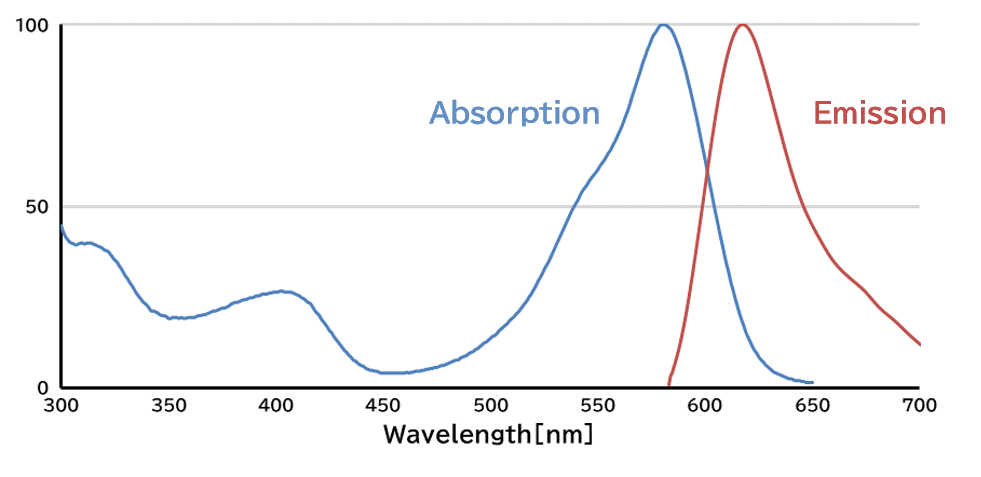
| Conjugate | SPICA Dye™ 594 (Excitation=575 nm, Emission=611 nm) |
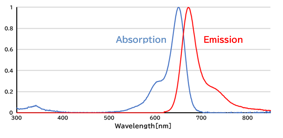
SPICA Dye™ 647 (Excitation=651 nm, Emission=670 nm) |
||
| Application | Immunohistochemistry (frozen section) 1:500-1,000 Immunocytochemistry 1:500-1,000 |
Immunohistochemistry (frozen section) 1:500-1,000 Immunocytochemistry 1:500-1,000 |
Immunohistochemistry (frozen section) 1:500-1,000 Immunocytochemistry 1:500-1,000 |
Immunohistochemistry (frozen section) 1:500-1,000 Immunocytochemistry 1:500-1,000 |
Excitation/Emission
(Excitation=575 nm, Emission=611 nm)
(Excitation=651 nm, Emission=670 nm)
Data
Performance Data
Photostability Evaluation Using Xenon Light Resistance Test Compared to Conventional Dyes
1) SPICA Dye™ 594
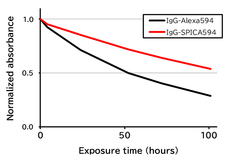
Using a xenon light resistance tester equipped with a 500 W xenon lamp and a glass filter, PBS solutions containing goat anti-rabbit IgG antibodies conjugated with either SPICA Dye™ 594 or Alexa Fluor® 594 (Jackson ImmunoResearch Inc., Product Number 111-005-003) were continuously subjected to xenon light irradiation. Changes in absorbance at the maximum absorption wavelength were plotted to compare the photostability of the two dyes.
[Result]
The SPICA Dye™ 594-conjugated antibody demonstrated greater photostability than the antibody conjugated with the competitor’s fluorescent dye.
2) SPICA Dye™ 647
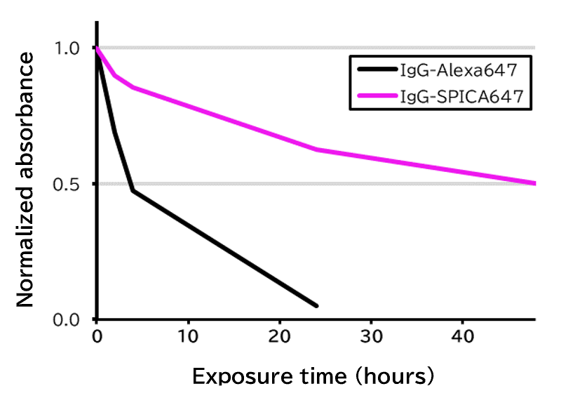
Using a xenon light resistance tester equipped with a 500 W xenon lamp and a glass filter, PBS solutions containing goat anti-rabbit IgG antibodies conjugated with either SPICA Dye™ 647 or Alexa Fluor® 647 (Jackson ImmunoResearch Inc., Product Number 111-005-003) were continuously subjected to xenon light irradiation. Changes in absorbance at the maximum absorption wavelength were plotted to compare the photostability of the two dyes.
[Result]
The SPICA Dye™ 647-conjugated antibody demonstrated greater photostability than the antibody conjugated with the competitor’s fluorescent dye.
Photostability Comparison in Immunohistochemistry
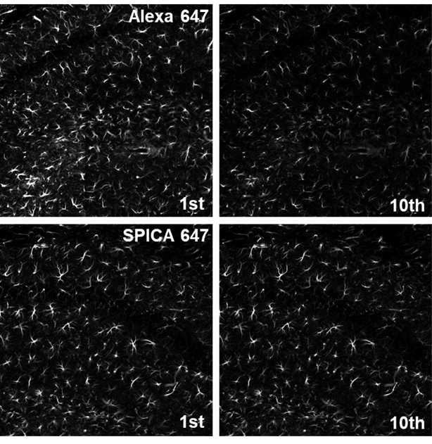
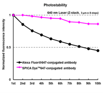
Immunostained mouse cryosections were subjected to 10 rounds of Z-stack acquisition using a confocal microscope, and fluorescence intensity was measured.
| Species | Mouse (C57BL6/J) |
|---|---|
| Site | Hippocampus |
| Sample | Frozen section |
| Primary Antibody | Anti GFAP, Monoclonal Antibody(MO389) (Product Number 018-27283) 1:500 |
| Secondary Antibody | Anti Mouse IgG, Goat, SPICA Dye™ 647 Conjugated (Product Number 010-28681) 1:500 Alexa Fluor® 647-AffiniPure Goat Anti-Mouse IgG (H+L) (Jackson ImmunoResearch Inc., Product Number 115-605-146) 1:500 |
| Microscope | CLSM (Nikon, ECLIPSE Ti) |
|---|---|
| Objective lens | Plan Apo VC ×20 (NA 0.75) |
| Excitation | 647 nm |
| Detection | 660-1,000 nm |
| Steps | 3.0 μm x 8 Steps |
| Scan Speed | 0.125 Frame/sec (pixel Dwell/5.3 μsec) |
[Result]
The SPICA Dye™ 647-conjugated antibody exhibited slower photobleaching than the Alexa Fluor® 647-conjugated antibody.
Photostability Evaluation by Widefield Microscopy
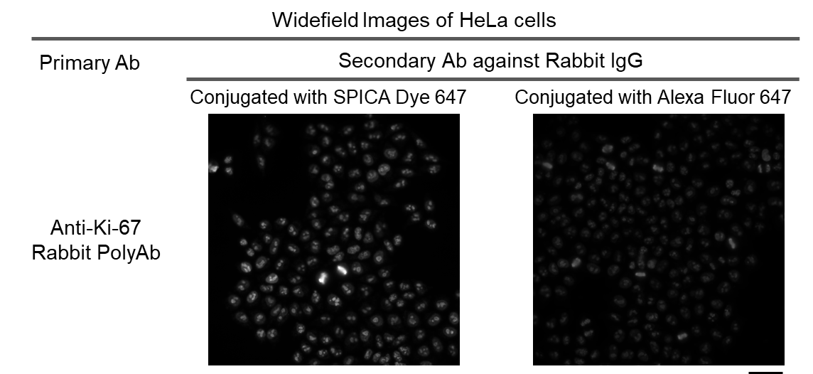

Photobleaching of near infrared fluorescent dyes for immunostaining Ki-67 in HeLa cells (widefield illumination).
(A) Representative images. Scale bar, 50 μm.
(B) Photobleaching curves.
(Alexa647_1-6); Goat anti-rabbit IgG (from Fuji*), Donkey anti-rabbit IgG (Jackson, 711-605-152), Goat anti-rabbit IgG (Thermo, A21244), Donkey anti-rabbit IgG (Thermo, A31573), Goat anti-rabit IgG (abcam, ab150083), Donkey anti-rabbit IgG (abcam, ab150075)
Microscope: IX83 P2ZF
Objective lens: UPLXAPO 40X/0.95 (NA=0.95) (Evident)
Ex/Em cube: Cy5-A-Basic (Semrock)
* Fuji: FUJIFILM Corp.
Photostability Evaluation by Confocal Microscopy
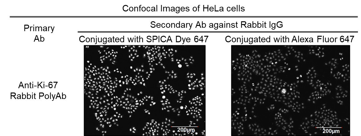
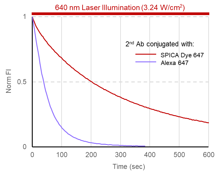
Photobleaching of near infrared fluorescent dyes for immunostaining Ki-67 in HeLa cells by confocal microscopy.
(A) Representative images.
Microscope: FV1200 CLSM
Objective lens: XLFLUOR 4X/340 (NA=0.28)
Illumination: 635 nm laser (0.81 W/cm2)
Scan Speed: 4.0 ms/pixel
Zoom: x 4.0
(B) Photobleaching curves.
Microscope: FV3000 CLSM (Evident)
Objective lens: UPLSAPO 40X(NA=0.95) (Evident)
Illumination: 640 nm laser (3.24 W/cm2)
Scan Speed: 2.0 ms/pixel
Zoom: x 2
* Alexa Fluor® is a registered trademark of Invitrogen Corporation.
Product List
- Open All
- Close All
SPICA Dye™ 594-conjugated Secondary Antibodies
SPICA Dye™ 647-conjugated Secondary Antibodies
For research use or further manufacturing use only. Not for use in diagnostic procedures.
Product content may differ from the actual image due to minor specification changes etc.
If the revision of product standards and packaging standards has been made, there is a case where the actual product specifications and images are different.
The prices are list prices in Japan.Please contact your local distributor for your retail price in your region.