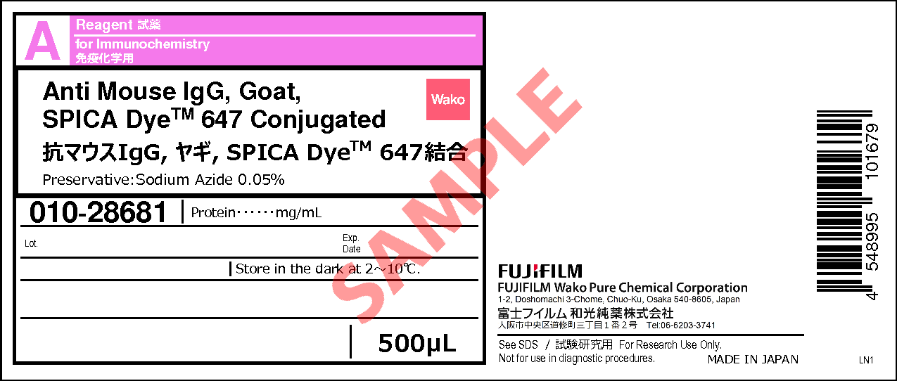
Anti Mouse IgG, Goat, SPICA DyeTM 647 Conjugated
- for Immunochemistry
- Manufacturer :
- FUJIFILM Wako Pure Chemical Corporation
- Storage Condition :
- Keep at 2-10 degrees C.
-
 Close
Close
- Structural Formula
- Label
- Packing
- SDS
|
Comparison
|
Product Number
|
Package Size
|
Price
|
Inventory
|
|
|---|---|---|---|---|---|
|
|
|
500uL
|
|
Please check here for notes on products and prices.
Document
Product Overview
Anti Mouse IgG, Goat, SPICA Dye™ 647 Conjugated is a secondary antibody conjugated with SPICA Dye™, a novel ultra-photostable fluorescent dye developed by Fujifilm. Compared to conventional fluorescent dyes, SPICA Dye™ is significantly more resistant to photobleaching. This antibody is suitable for both immunohistochemistry and immunocytochemistry.
Antibody Information
| Clonality | Polyclonal |
|---|---|
| Antigen | Mouse IgG |
| Host | Goat |
| Formulation | PBS, 0.05% Sodium azide |
| Conjugate | SPICA Dye™ 647 |
| Cross-reactivity | Mouse |
| Application | Immunohistochemistry (frozen section) 1:500-1,000 Immunocytochemistry 1:500-1,000 |
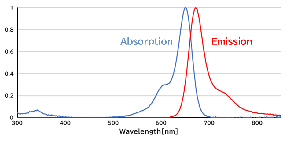
Excitation/Emission
SPICA Dye™ 647
Protocol (Example)
Immunohistochemistry of Mouse Brain Sections using Anti-GFAP Antibody and Anti Mouse IgG, Goat, SPICA Dye™ 647 Conjugated
- 1.Perfusion fixation
- Perfuse with 0.1% sodium citrate/PBS to remove blood, followed by perfusion with 4% PFA.
- 2.Post-fixation
- 4% PFA (24 hours)
- 3.Immersion in sucrose
- 30% Sucrose/PBS (4 °C, from overnight to approximately 2 days)
- 4.Preparation of frozen sections
- After removing sucrose, prepare frozen blocks and cut 30 μm thick sections using a cryostat.
- 5.Blocking
- 5% BSA and 2% normal donkey serum in 0.3% Triton X-100/PBS (RT, 2 hours)
- 6.Primary antibody reaction
- Anti GFAP, Monoclonal Antibody(MO389) (Product Number 018-27283) 1:1,000(4 °C, Overnight)
- 7.Washing
- 0.3% TritonX-100/PBS (5 minutes x 3)
- 8.Secondary antibody reaction
- Anti Mouse IgG, Goat, SPICA Dye™ 647 Conjugated (This product) 1:500 in 0.3% Triton X-100/PBS (RT, 2 hours)
- 9.Washing
- 0.3% TritonX-100/PBS (5 minutes x 3)
- 10.Mounting
- The sample is sealed in mounting medium (ex. Invitrogen™ ProLong Glass) and stored at 4 °C in a dark place.
Data
Performance Data
Photostability Evaluation Using Xenon Light Resistance Test Compared to Conventional Dyes
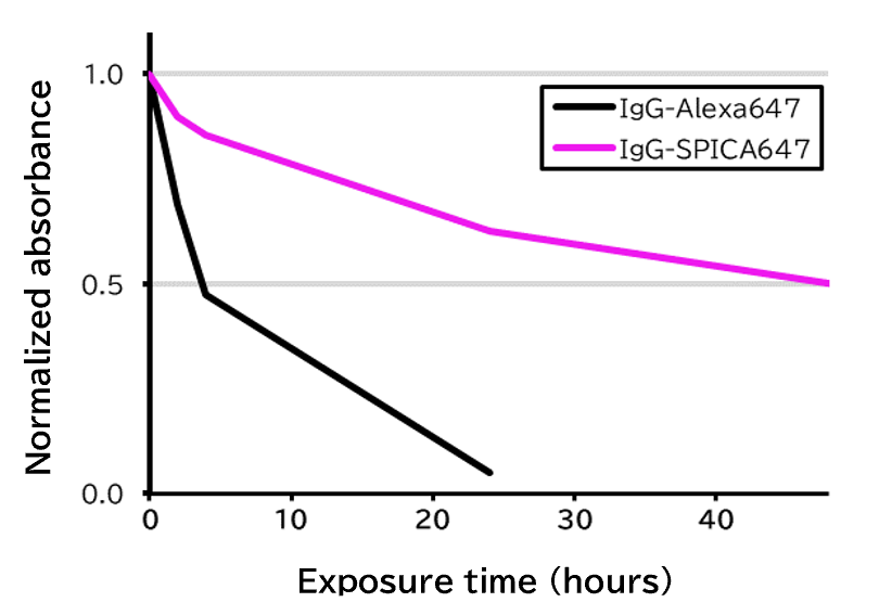
Using a xenon light resistance tester equipped with a 500 W xenon lamp and a glass filter, PBS solutions containing goat anti-rabbit IgG antibodies conjugated with either SPICA Dye™ 647 or Alexa Fluor® 647 (Jackson ImmunoResearch Inc., Product Number 111-005-003) were continuously subjected to xenon light irradiation. Changes in absorbance at the maximum absorption wavelength were plotted to compare the photostability of the two dyes.
[Result]
The SPICA Dye™ 647-conjugated antibody demonstrated greater photostability than the antibody conjugated with the competitor’s fluorescent dye.
Photostability Comparison in Immunohistochemistry
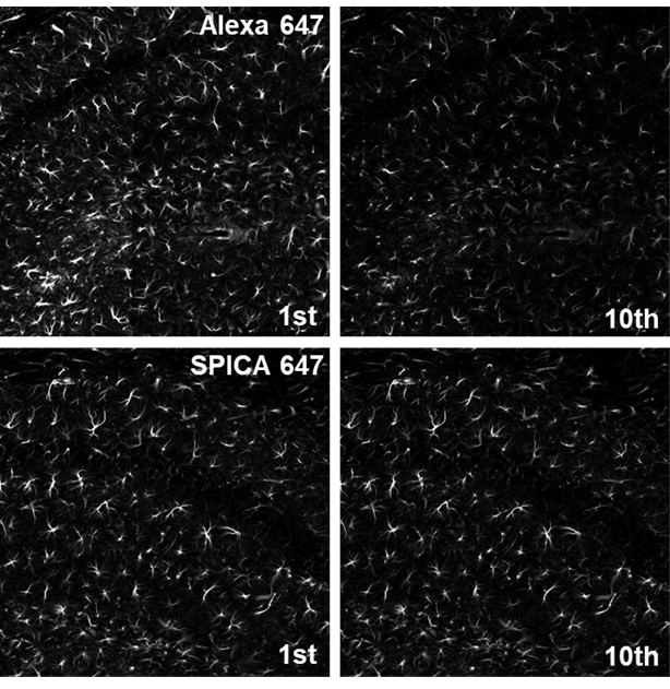
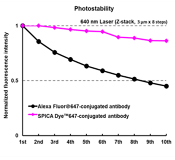
Immunostained mouse cryosections were subjected to 10 rounds of Z-stack acquisition using a confocal microscope, and fluorescence intensity was measured.
| Species | Mouse (C57BL6/J) |
|---|---|
| Site | Hippocampus |
| Sample | Frozen section |
| Primary Antibody | Anti GFAP, Monoclonal Antibody(MO389) (Product Number 018-27283) 1:500 |
| Secondary Antibody | Anti Mouse IgG, Goat, SPICA Dye™ 647 Conjugated (This product) 1:500 Alexa Fluor® 647-AffiniPure Goat Anti-Mouse IgG (H+L) (Jackson ImmunoResearch Inc., Product Number 115-605-146) 1:500 |
| Microscope | CLSM (Nikon, ECLIPSE Ti) |
|---|---|
| Objective lens | Plan Apo VC ×20 (NA 0.75) |
| Excitation | 647 nm |
| Detection | 660-1,000 nm |
| Steps | 3.0 μm x 8 Steps |
| Scan Speed | 0.125 Frame/sec (pixel Dwell/5.3 μsec) |
[Result]
The SPICA Dye™ 647-conjugated antibody exhibited slower photobleaching than the Alexa Fluor® 647-conjugated antibody.
* Alexa Fluor® is a registered trademark of Invitrogen Corporation.
FAQ
About antibody
- How is this antibody purified?
- This antibody is purified by antigen affinity chromatography.
- What is the concentration of the antibody?
- 1.4-1.6 mg/mL (as a protein concentration). The actual concentration is indicated on the label.
Overview / Applications
| Outline | Secondary antibodies are versatile and can be used in a variety of detection systems, including immunostaining. Anti-Mouse IgG secondary antibodies are affinity-purified antibodies with characterized specificity for mouse IgG and are useful in the detection or sorting of its target. Anti Mouse IgG, Goat, SPICA DyeTM 647 Conjugated is conjugated to SPICA DyeTM 647 fluorescent dye under optimal conditions. Since this product is a polyclonal antibody and multiple secondary antibodies can bind to one primary antibody, signal amplification can also be expected. SPICA DyeTM 647 (Excitation:651 nm, Emission:670 nm) has superior light resistance compared to existing fluorescent dyes and is useful for experiments such as transparency and ultraresolution microscopy. [Class] Goat IgG [Species cross reactivity] Mouse [Application] Immunocytochemistry 1:500 - 1:1,000 Immunohistochemistry (frozen section) 1:500 - 1:1,000 Optimal concentration should be determined by each laboratory for each application. |
|---|
Property
| Appearance | Liquid |
|---|---|
| Concentration | Described on the label Protein : 1.4 - 1.6mg/mL |
Manufacturer Information
Alias
For research use or further manufacturing use only. Not for use in diagnostic procedures.
Product content may differ from the actual image due to minor specification changes etc.
If the revision of product standards and packaging standards has been made, there is a case where the actual product specifications and images are different.
The prices are list prices in Japan.Please contact your local distributor for your retail price in your region.