StemSure®Serum Replacement
StemSure® Serum Replacement is a serum replacement for ES/iPS cell culture. Since serum contains factors that induce differentiation of cells, serum replacements are commonly used when culturing ES/iPS cells to maintain their undifferentiated states. ES/ iPS cells can thus be cultured safely using this product.
The product is safe to handle as it is not categorized as a poisonous substance. Murine ES cells cultured in medium containing StemSure Serum Replacement have been used to create chimera mice and to achieve germline transmission.
Features
- Can be used for chimera formation and germline transmission.
- Can be used as a serum replacement for culturing murine ES cells and human iPS cells.
- Not a poisonous substance.
Validation Tests
- Colony formation assay (using murine ES cell line D3)
- Alkaline phosphatase assay (using murine ES cell line D3)
- Sterility test
- pH
- Osmotic pressure
- Endotoxin test
- Mycoplasma test
Establishing chimera mice and germline transmission
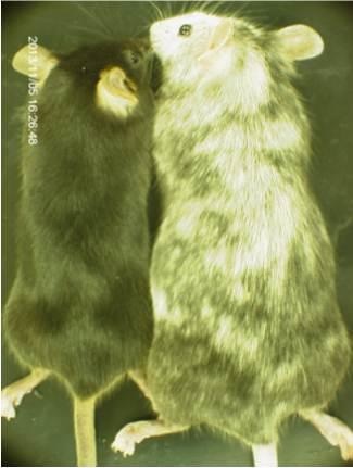
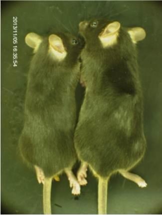
A targeting vector constructed with HaloTag® cDNA was transduced into murine ES cells, and the cells were cultured in a medium containing StemSure® Serum Replacement (SSR). After confirming successful transduction, the murine ES cells were injected into blastocysts of host ICR mice and transplanted into the uterus of ICR mice. The resulting chimera mice and C57BL/6J mice were crossed to generate F1 mice.
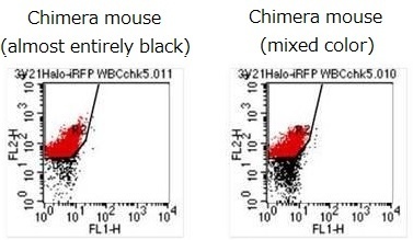
Chimera mice
Blood cells were collected from the chimera mice, and HaloTag® proteins were stained with tetramethylrhodamine. Chimera formation was confirmed by the presence of positively-stained blood cells expressing murine ES-derived HaloTag®.
F1 mice
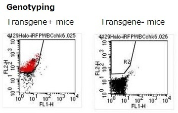
HaloTag® was expressed in all blood cells in some of the F1 mice. This suggested that some mice were generated entirely from ES cells cultured in the medium containing SSR.
<Data source: Dr. Y Miwa, Laboratory Animal Resource Center, University of Tsukuba. >
Halo Tag® is a registered trademark of Promega Corp.
Culturing human iPS cell line 201B7
Colony morphology and ALP staining
StemSure® Serum Replacement (SSR) was used to culture human iPS cell line 201B7. These cells stained positive for ALP.
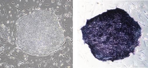
<Culture medium>
D-MEM/Ham's F-12 + 20% SSR + 2mmol/l L-Glutamine +1×MEM Non-essential AminoAcids + 0.1mmol/l StemSure® 2-Mercaptoethanol + 1×Penicillin-Streptomycin + 5ng/ml bFGF
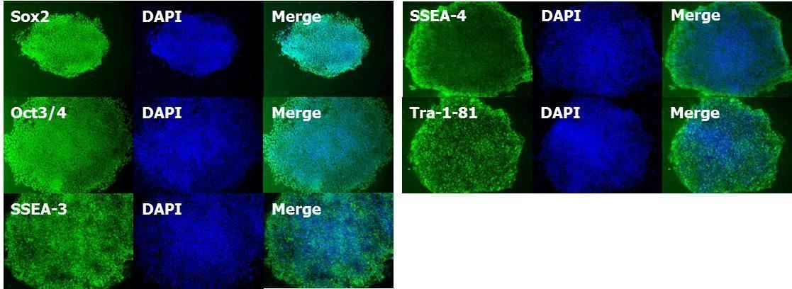
Identifying expression of markers for undifferentiated cells
Huma iPS cell line 201B7 was cultured using SSR. Cells stained positive for markers of undifferentiated cells (Sox2, Oct3/4, SSEA-3, SSEA-4, Tra-1-81).
Culturing Murine ES Cell Line D3
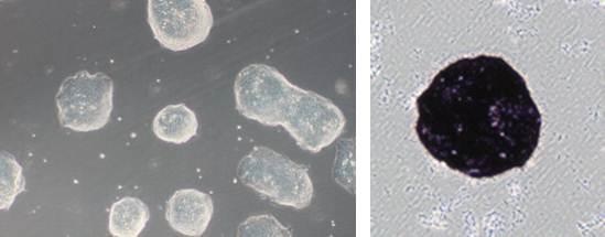
Cell morphology and ALP staining
StemSure® Serum series were used to culture murine ES cell line D3. Colonies of these cells appeared shiny, which is characteristic of murine ES cells. The cells stained positive for ALP.
Cell growth curve
Murine ES cell line D3 was cultured in either SS-DMEM + SSR + 2ME or DMEM (from Company A) + serum-equivalent + 2ME for 14 passages to compare cell growth. As shown below, cells grown in StemSure® series had the equivalent growth curve as those grown in a medium containing the product from Company A.
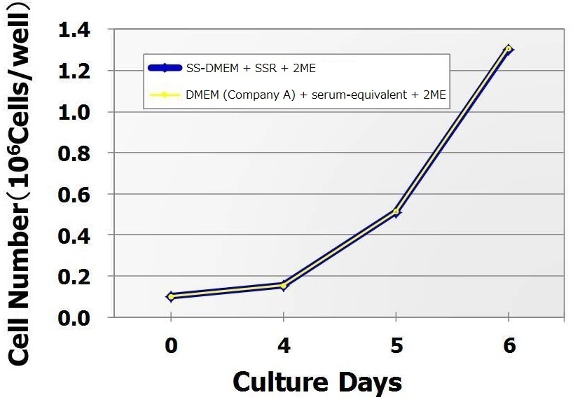
< Culture medium for StemSure® >
StemSure® D-MEM + 15% SSR + 2mmol/l L-Glutamine + 1×MEM Non-essential Amino Acids + 0.1mmol/l StemSure® 2-Mercaptoethanol + 1×Penicillin-Streptomycin + 1,000units/ml StemSure® LIF
(Using collagen-coated 12-well plates)
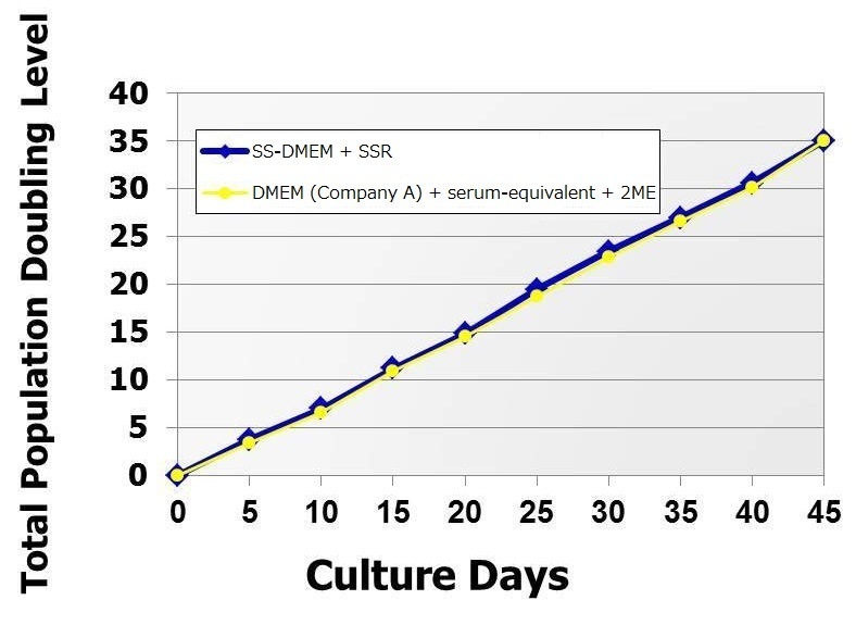
Cell doubling time
Murine ES cell line D3 was passaged in the two culture media, and the association between the duration of cell culture and doubling time was examined. As shown below, cells grown in StemSure® series had the equivalent doubling time as those grown in a medium containing the product from Company A.
< Culture medium >
StemSure® D-MEM + 15% SSR + 2mmol/l L-Glutamine + 1×MEM Non-essential Amino Acids + 0.1mmol/l StemSure® 2-Mercaptoethanol + 1×Penicillin-Streptomycin + 1,000units/ml StemSure® LIF
(Using collagen-coated 12-well plates)
Identifying expression of markers for undifferentiated cells
Murine ES cell line D3 was cultured using the StemSure® series. After 8 passages, cells stained positive for markers of undifferentiated cells (Nanog, Oct3/4, Sox2, SSEA-1).

< Culture medium >
StemSure® D-MEM + 15% SSR + 2mmol/l L-Glutamine + 1×MEM Non-essential Amino Acids + 0.1mmol/l StemSure® 2-Mercaptoethanol+ 1×Penicillin-Streptomycin + 1,000units/ml StemSure® LIF
Identifying teratoma formation
Murine ES cell line D3 was cultured for multiple passages using the StemSure® series. Cells were then injected subcutaneously in immunocompromised mice. Teratomas formed subcutaneously, and various tissue types including nerve tissue (derived from ectoderm), cartilage tissue (derived from mesoderm), and luminal structures with ciliated epithelium (derived from endoderm) were found in the teratomas.

< Culture medium >
StemSure® D-MEM + 15% SSR + 2mmol/l L-Glutamine + 1×MEM Non-essential Amino Acids + 0.1mmol/l StemSure® 2-Mercaptoethanol + 1×Penicillin-Streptomycin + 1,000units/ml StemSure® LIF
Product List
- Open All
- Close All
For research use or further manufacturing use only. Not for use in diagnostic procedures.
Product content may differ from the actual image due to minor specification changes etc.
If the revision of product standards and packaging standards has been made, there is a case where the actual product specifications and images are different.


