Anti Laminin Antibodies
Laminin is a glycoprotein that constitutes the extracellular matrix (ECM) and plays an important role in cell adhesion, migration, and proliferation. In particular, laminin, together with type IV collagen, is a major component of the basement membrane. Fujifilm Wako offers guinea pig anti-laminin antibodies that are useful for multiplex staining.
What is Laminin?

Basic Structure of Laminin
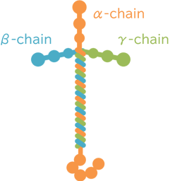
Laminin is a glycoprotein with a molecular weight of approximately 900 kDa. It consists of three chains-α, β, and γ-that assemble into a cross-shaped structure. Each chain has multiple isoforms, and laminin types are named based on the combination of isoforms (for example, laminin composed of α1/β1/γ1 is referred to as laminin-111).
Laminin is well-known as a key component of the extracellular matrix (ECM) and plays an important role in cell adhesion, migration, and proliferation. In particular, laminin, together with type IV collagen, is considered a major component of the basement membrane. In the central nervous system, laminin is almost exclusively expressed in the basement membranes surrounding blood vessels and is used as a marker for these vessels. Previous studies have shown that the inner (endothelial) layer of the basement membrane in blood vessels contains laminin-411 and -511, while the outer (parenchymal) layer contains laminin-211 and -1111). Laminin also plays a critical role in the formation and function of the blood-brain barrier (BBB).
Anti Laminin, Guinea Pig
The “Anti Laminin , Guinea Pig” is a guinea pig polyclonal antibody, raised against Laminin2-6). It can be used to perform multiplex immunohistochemistry.
Antibody Information
| Clonality | Polyclonal |
|---|---|
| Antigen | Laminin isolated from Engelbreth-Holm-Swarm murine sarcoma basement membrane |
| Host | Guinea pig |
| Formulation | Antiserum diluted in PBS |
| Conjugate | Unconjugated |
| Cross-reactivity | Mouse, Rat |
| Application | Immunohistochemistry (Frozen Section) 1:500 * 6 hours of post-fixation is recommended |
Application Data
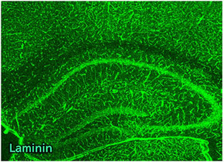
Immunohistochemistry

Species: Mouse
Site: Hippocampus
Sample: Frozen section
Antibody concentration: 1:500
Data by courtesy of
Dr. Miyata, Department of Applied Biology, Kyoto Institute of Technology

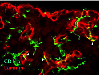
Species: Mouse
Site: Medulla oblongata (Area postrema)
Sample: Frozen section
Antibody concentration: 1:500
Data by courtesy of
Dr. Miyata, Department of Applied Biology, Kyoto Institute of Technology
References
- Halder, S. K. et al.: Neural Regen. Res., 18(12), 2557(2023).
The importance of laminin at the blood-brain barrier - Morita, S. et al.: Cell Tissue Res., 363, 497(2016).
Heterogeneous vascular permeability and alternative diffusion barrier in sensory circumventricular organs of adult mouse brain - Nishikawa, K. et al.: J. Neuroendocrinol., 29(2), (2017).
Structural Reconstruction of the Perivascular Space in the Adult Mouse Neurohypophysis During an Osmotic Stimulation - Takagi, S. et al.: J. Neuroimmunol., 331, 74(2019).
Microglia are continuously activated in the circumventricular organs of mouse brain - Muneoka, S. et al.: J. Neuroimmunol., 331, 58(2019).
TLR4 in circumventricular neural stem cells is a negative regulator for thermogenic pathways in the mouse brain - Murayama, S. et al.: J. Neuroimmunol., 334, 576973(2019).
Activation of microglia and macrophages in the circumventricular organs of the mouse brain during TLR2-induced fever and sickness responses
Product List
- Open All
- Close All
Anti Laminin, Guinea Pig (Polyclonal Antibody)
For research use or further manufacturing use only. Not for use in diagnostic procedures.
Product content may differ from the actual image due to minor specification changes etc.
If the revision of product standards and packaging standards has been made, there is a case where the actual product specifications and images are different.


