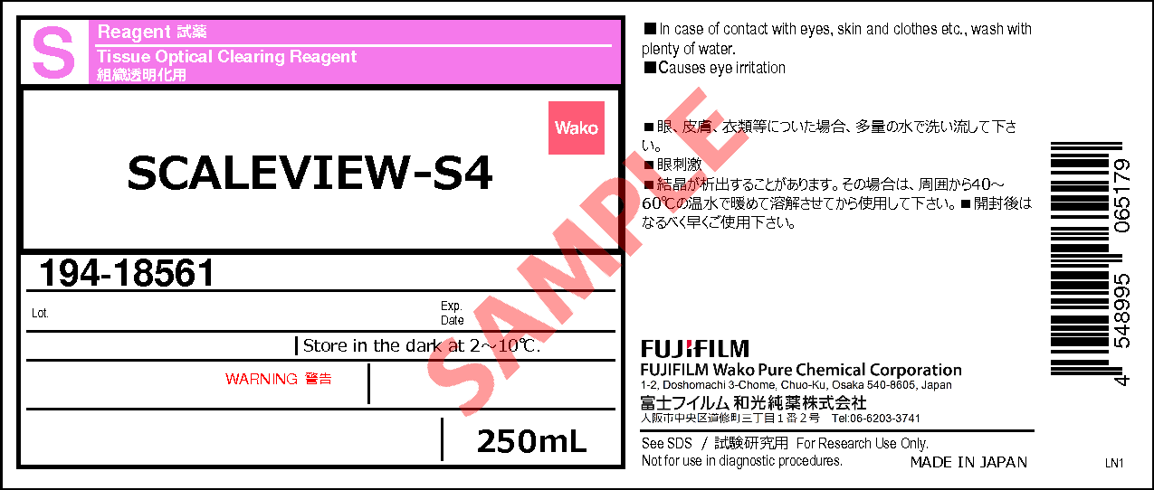
SCALEVIEW-S4
- Tissue Optical Clearing Reagent
- Manufacturer :
- FUJIFILM Wako Pure Chemical Corporation
- Storage Condition :
- Keep at 2-10 degrees C.
- GHS :
-


-
 Close
Close
- Structural Formula
- Label
- Packing
- SDS
|
Comparison
|
Product Number
|
Package Size
|
Price
|
Inventory
|
|
|---|---|---|---|---|---|
|
|
|
250mL
|
|
In stock in Japan |
Please check here for notes on products and prices.
Document
Product Overwiew
SCALEVIEW-S4 is a reagent included in the SCALEVIEW-S Trial Kit, which is used in the Scale-S tissue clearing method. In addition to being used as part of the SCALEVIEW-S Trial Kit protocol, an optional simplified protocol allows spheroids and organoids to be cleared simply by immersing them in SCALEVIEW-S4.
Simplified Clearing Protocol for Spheroids/Organoids Using SCALEVIEW-S4 (Optional)
The following is a simplified method for clearing spheroids and organoids using only SCALEVIEW-S4. While this protocol offers ease of use, its clearing efficiency may be slightly lower than that of the SCALEVIEW-S Trial Kit. It is recommended for small-sized spheroids/organoids (approximately, a few hundred µm thick). For clearing spheroids/organoids using the SCALEVIEW-S Trial Kit, please refer to the product overview page of the SCALEVIEW series. Additionally, if spheroids tend to float, the combined use of SCALEVIEW-S0 and SCALEVIEW-A2 is recommended.
Features
- Enabling spheroids and organoids clearing
- Clears spheroids and organoids in about one day1)
- Compatible with Immunostaining and fluorescent dyes staining
Samples suitable for clearing
Spheroids, Organoids, Neurosphere
> Reagents
> Instruments
- Plate for spheroid formation (PrimeSurface 96 Slit-well Plate (Manufacturer Product No. MS-9096S) is convenient.)
- Observation plate (We recommend those with a cover glass on the bottom.)
- SCALEVIEW-S4 fluorescence microscope and recommended objective lens ※1 for clearing specimens
※1 Ensure that the objective lenses are compatible with the refractive indices (RI=1.49).
Consulting the microscope manufacturer before data acquisition is recommended.
Protocol
| Fixation | Clearing | Observation※2 |
|---|---|---|
| 4%PFA/PBS (-) | SCALEVIEW-S4 | SCALEVIEW-S4 |
| RT 1 hour |
37°C 4 hours |
RT |
※2 Immobilizing the samples by embedding them in 1.5% (w/v) agarose facilitates observation.
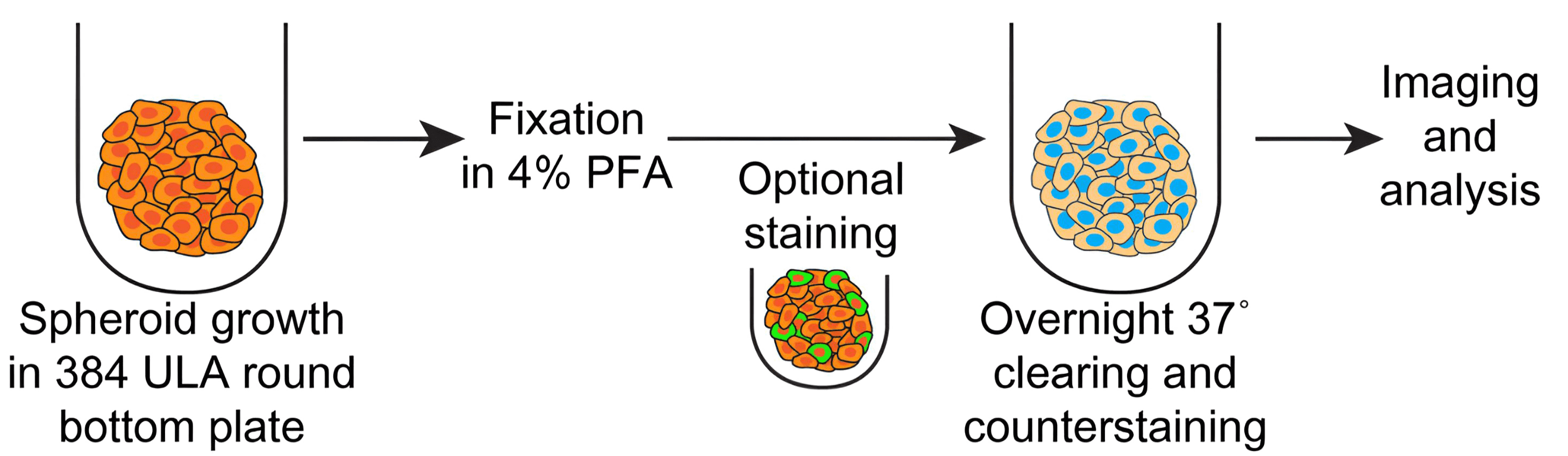
Data (Clearing of Spheroids/Organoids)
The following data represent samples cleared using SCALEVIEW-S4 alone. For spheroid/organoid clearing with the SCALEVIEW-S Trial Kit, please refer to the product overview page of the SCALEVIEW series.
Spheroids
Clearing of T47D cell Spheroids
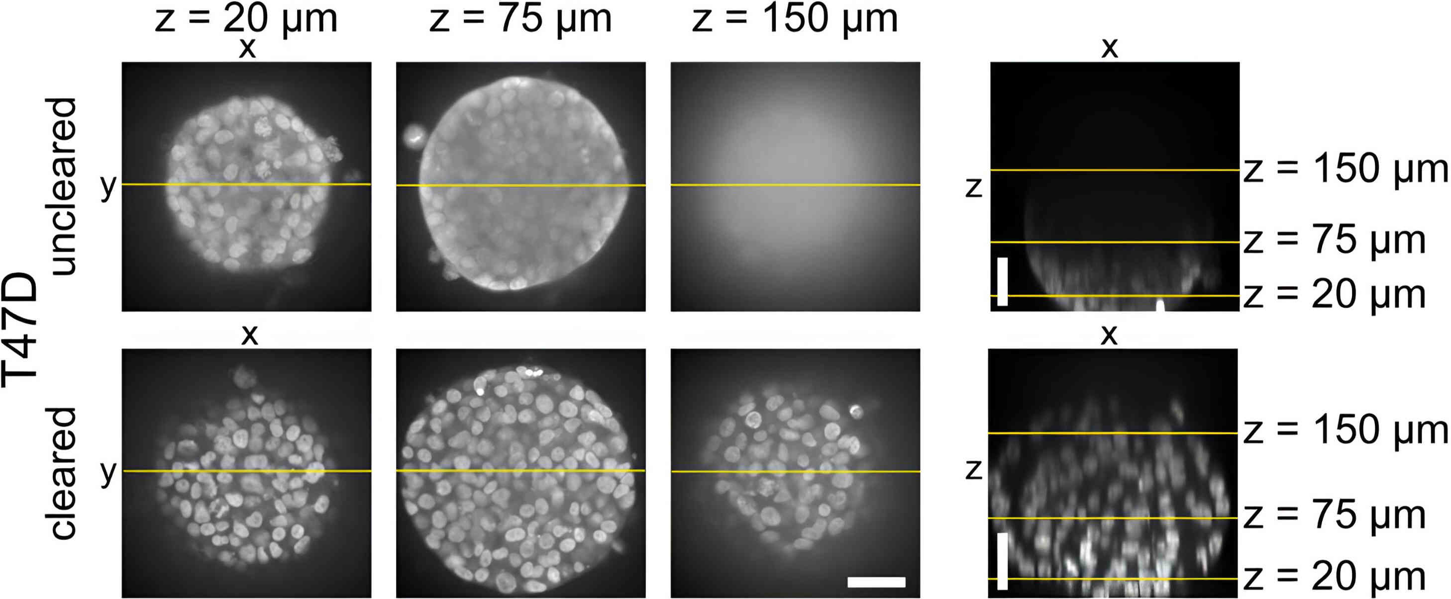
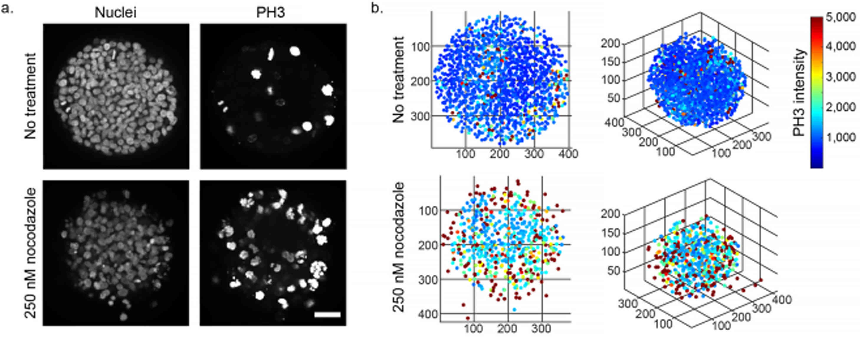
Clearing with SCALEVIEW-S4 enables visualization of the drug’s effects on both the surface and interior of the spheroid. Reproduced from reference 1) under CCBY 4.0.
Organoids
Clearing of F-PDO® (Cultured cells derivered from cancer tissue)
RLUN14 (Carcinoma in situ of the intraepithelial gland/Bronchioalveolar carcinoma)
- Microscope:
- Confocal microscope (Evident, FV3000, Inverted)
- Objective lens:
- UPLSAPO30 x S
- Antibody:
- ZO-1-Alexa488 conjugate
- Dyes:
- Actistain670 (Phalloidin)DAPI
RLUN20 (Colloid gland cancer)
- Microscope:
- Confocal microscope (Evident, FV3000, Inverted)
- Objective lens:
- UPLSAPO30 x S
- Antibody:
- ZO-1-Alexa488 conjugate
- Dyes:
- Actistain670 (Phalloidin)DAPI
Reference
- Molly E. B. et al.: Sci. Rep., 8, 11135(2018).
A high-throughput imaging and nuclear segmentation analysis protocol for cleared 3D culture models
Overview / Applications
| Outline | SCALEVIEW-S4 is a tissue clearing reagent designed for use in the ScaleS, AbScale, AbScale-G (three-dimensional antibody staining method), and ChemScale (three-dimensional fluorescent dye staining method). SCALEVIEW-S4 is a solution specifically designed for refractive index adjustment and mounting of biological tissues, with a refractive index set at approximately 1.47. Additionally, a simplified protocol has been reported, in which simply immersing spheroids or organoids in SCALEVIEW-S4 allows for effective tissue clearing. To perform the AbScale or ChemScale, this product must be used in conjunction with SCALEVIEW-S0 (Product Number: 196-18521), SCALEVIEW-A2 (Product Number: 193-18455) ans deScale Solution (Product Number: 041-34425). Additionally, the AbScale-G method requires the AbScale Solution Kit (Product Number: 293-97001). For detailed protocols, data, and further information, please visit our website via the link below. https://labchem-wako.fujifilm.com/us/category/00593.html |
|---|
Property
| Appearance | Colorless - nearly colorless, clear - nearly clear liquid |
|---|
Manufacturer Information
Alias
For research use or further manufacturing use only. Not for use in diagnostic procedures.
Product content may differ from the actual image due to minor specification changes etc.
If the revision of product standards and packaging standards has been made, there is a case where the actual product specifications and images are different.
The prices are list prices in Japan.Please contact your local distributor for your retail price in your region.