Fluorescent Probes (Organelle)
Some fluorescent dyes tend to accumulate in specific locations in the cell. For example, the lipophilic fluorescent dye rhodamine penetrates the plasma membrane and accumulates in mitochondria. This is because rhodamine-type fluorescent dyes have a cationic backbone and are electrostatically attracted to mitochondria, which have a negative membrane potential. Thus, specific detection of organelles is possible by taking advantage of the properties of fluorescent dyes. Fujifilm Wako offers fluorescent probes for each organelle and for visualizing changes in membrane potential.
Product Line-up
More Information
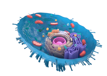
Fluorescent Probes for Labeling Organelles
Specific organelles can be selectively stained by utilizing their physical and chemical properties.
The fluorescent probes used for labeling each organelle and their properties are summarized below.
Nucleus
The nucleus can be stained by using fluorescent dyes that specifically bind to DNA. Typical staining reagents include DAPI, Hoechst 33258, and Hoechst 33342. The fluorescence intensity of these fluorescent dyes increases when they bind to the minor groove of double-stranded DNA, enabling specific staining of double-stranded DNA. Although these reagents can be used for nuclear staining of both living and dead cells, Hoechst 33342 is often used for living cells due to its excellent cell permeability, and DAPI is often used for dead cells due to its high fluorescence intensity.
The nucleolus is a structure within the nucleus with no membranes and is responsible for the biosynthesis of ribosomes. Since the nucleolus is enriched with ribosomal RNA, a constituent of ribosomes, intense staining can be achieved with a fluorescent dye that binds to RNA.
Mitochondria
Mitochondria are energy-producing organelles. The membrane potential is formed in mitochondria by the proton gradient generated by redox reactions, and staining is performed with fluorescent dyes that accumulate in mitochondria in a membrane potential-dependent manner.
Mitochondrial staining reagents include rhodamine 123, tetramethylrhodamine methyl ester (TMRE), and JC-1. Rhodamine has a cationic backbone and is electrostatically attracted to mitochondria with negative membrane potential. JC-1 emits red fluorescence in normal mitochondria, but when the mitochondrial membrane potential decreases, the aggregation of the dye weakens, and the fluorescence wavelength changes to green. This property can be used to measure mitochondrial membrane potential.
Endoplasmic Reticulum and Golgi Apparatus
The endoplasmic reticulum is a reticular membrane system in the cytoplasm and is the site of synthesis of membrane proteins and secretory proteins. Folding of newly synthesized proteins also takes place in the endoplasmic reticulum. Proteins synthesized in the endoplasmic reticulum undergo glycosylation in the Golgi apparatus and are delivered to various locations, including the extracellular space.
Fluorescently labeled lipids are used for staining the endoplasmic reticulum and Golgi apparatus. Ceramide is known to selectively accumulate in the Golgi apparatus. Fluorescently labeled analogues of ceramide and sphingomyelin are used as fluorescent probes.
References
"How to select and use fluorescent and luminescent reagents for successful experiments” ed. by Miwa, Y., Yodosha, Japan, (2007). (Japanese).
For research use or further manufacturing use only. Not for use in diagnostic procedures.
Product content may differ from the actual image due to minor specification changes etc.
If the revision of product standards and packaging standards has been made, there is a case where the actual product specifications and images are different.
The prices are list prices in Japan.Please contact your local distributor for your retail price in your region.