Fluorescent Probes (Neuroscience)
Visualizing neurons and neural circuits help us to understand the part of mechanism how neurons transmit information. Anterograde or retrograde tracers are often used to label of neurons and their circuits. In addition, improvements in observation technology such as microscopes have made it possible to observe fine structures such as synapses. Fujifilm Wako has a line up of neuronal tracers, synaptic observation fluorescent probes, and selective fluorescent probes for senile plaques and neurofibrillary tangles.
More Information
Fluorescent Probes Used for Imaging Neurons and Neuronal Circuits
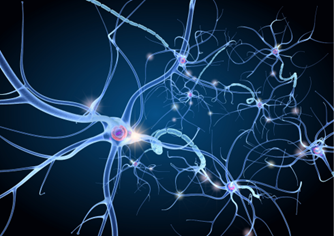
Neuronal tracers are generally used for imaging neurons and neuronal circuits. Radioactive amino acids and peroxidase (HRP) were used in the past, but now fluorescent dyes are used as tracers. Neuronal tracers are classified into anterograde and retrograde types. As for anterograde tracers, neurons are labeled by injecting dyes into the neuronal cell bodies and letting the dyes travel through the anterograde axoplasmic flow from the cell body to the nerve terminal, while for retrograde tracers, neurons are labeled by injecting dyes into the nerve terminals and letting the dyes travel through the retrograde axoplasmic flow from the nerve terminal to the cell body.
Carbocyanine dyes are widely used as neuronal tracers, because of their stable and strong fluorescence in lipids such as cell membranes. Although they can be used as both anterograde and retrograde tracers in juvenile animals, anterograde labeling is known to be difficult in adults due to advanced myelination of the nervous system. Fujifilm Wako's DiIC18(3) is a type of carbocyanine dye with an excitation wavelength of 548 nm and a fluorescence wavelength of 565 nm. In addition, Fluoro-Gold (retrograde tracer) and labeled dextran (anterograde or retrograde tracer) are also available as neuronal tracers.
Lucifer yellow, a hydrazine derivative, is also a fluorescent dye used for imaging neurons. When strong blue excitation light is applied to neurons that have been injected with lucifer yellow in the presence of diaminobenzidine (DAB), DAB turns black through a photooxidation reaction. This method, called photo conversion, is known to visualize neurons and enables electron microscopic observation.
Fluorescent Probes Used for Imaging synapses
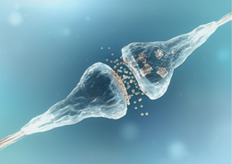
At synapses, exocytosis and endocytosis are frequently repeated for neurotransmitters release and vesicles recycling. Dyes that can label endocytic vesicles are therefore used for imaging synapses. Fluorescent cationic styryl dyes have been used for imaging synapses as fluorescent dyes that can label endocytic vesicles. They have a highly hydrophilic cationic charge head group at one end and a lipophilic tail at the other end. Although they are nonfluorescent in aqueous solutions, they exhibit strong fluorescence when bound to lipids such as cell membranes. As the dyes are released from vesicles along with neurotransmitters and cause a decrease in the fluorescent signal, the change in fluorescence intensity is thought to reflect the amount of endocytosis/exocytosis or synaptic activity.
A representative fluorescent cationic styryl dye is FM®* dye, developed by Fei Mao et al. ViVidFluor from Fujifilm Wako, is equivalent to FM® dye.
* FM® is a registered trademark of Life Technologies.
References
1) "How to select and use fluorescent and luminescent reagents for successful experiments” ed. by Miwa, Y., Yodosha, Japan, (2007). (Japanese).
For research use or further manufacturing use only. Not for use in diagnostic procedures.
Product content may differ from the actual image due to minor specification changes etc.
If the revision of product standards and packaging standards has been made, there is a case where the actual product specifications and images are different.
The prices are list prices in Japan.Please contact your local distributor for your retail price in your region.