AkaLumine-HCl (TokeOni)
AkaLumine-HCl is a luminescent substrate of luciferase used for in vivo bioluminescence imaging. This product has a wavelength at the near-infrared region (670-680nm) that less susceptible to absorption by water and hemoglobin than luciferin. Therefore, it's possible to observe more deep inside the body.
An example of use is an artificial bioluminescence system called AkaBLI, developed by Dr. Atsushi Miyawaki and Dr. Satoshi Iwano, etc.
This method uses Akaluc as luciferase and AkaLumine-HCl as luciferin. It's possible to detect deep tissues noninvasively at the single-cell level and track cell activity over time in the same living animals.
Overview
AkaLumine-HCl is a luciferin analog that has a luminescence peak at 670 ~ 680 nm. The peak range is in the near-infrared (NIR) window (also known as the optical window). Since the adsorption of hemoglobin and water is small in the optical window, Aka Lumine is well suited to in vivo imaging.
Features
- Shows wavelength at near-infrared region (670-680nm)
- Less susceptible to absorb by water and hemoglobin
- Suitable for deep imaging
- Well soluble in water and saline
Structural formula / Emission spectrum
Structure formula
Fig1. AkaLumine-HCl
Luminescence Wavelength
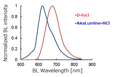
Fig2. Bioluminescence emission spectra of d-luciferin (d-luci) and AkaLumine-HCl (Aka-HCl).
Observation examples of AkaLumine-HCl
| Data provided by Dr. Takahiro Kuchimaru and Dr. Shinae Kizaka-Kondoh, School of Life Science and Technology, Tokyo Institute of Technology. Dr. Shojiro Maki, Graduate School of Informatics and Engineering, The University of Electro-Communications |
in vivo imaging data using mouse
Mice with LLC/Firefly luciferase (luc) subcutaneous tumor were administered intraperitoneally D-luciferin and AkaLumine-HCl at two substrate concentrations. After 15 minutes, luminescent imaging was got and analyzed quantitatively. As a result, it was found that when AkaLumine-HCl was used as a substrate, the substrate concentration did not easily affect the emission intensity.
In this experiment, AkaLumine-HCl was administrated to the same mouse 4 hours after D-luciferin administration.
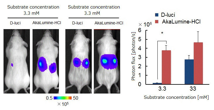
(Fig.3) Left: luminescent images 15 min after intraperitoneal administration
Right: quantitative analysis of emission intensity (n=4 *P<0.05 (t-test))
in vitro imaging data using cells
Mouse lung cancer cells constitutively expressing luc (LLC/luc) were added D-luciferin and AkaLumine-HCl at various substrate concentrations. And, luminescent imaging was got and analyzed quantitatively. As a result, it was found that when AkaLumine-HCl was used as a substrate, the substrate concentration did not easily affect the emission intensity.
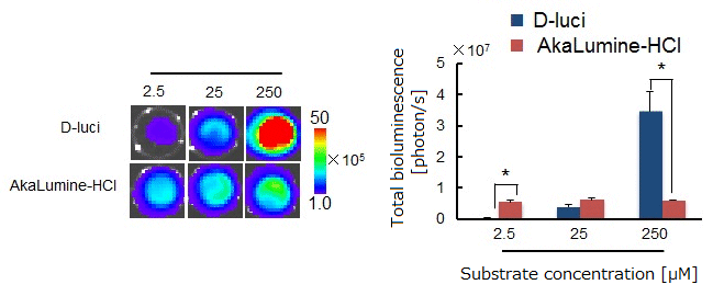
(Fig.4) Left: luminescent imaging after administration
Right: quantitative analysis of emission intensity (n=3 *P<0.05 (t-test))
Bioluminescence images using AkaBLI system
| Data provided by Dr. Satoshi Iwano and Dr. Atsushi Miyawaki, Laboratory for Cell Function Dynamics, Brain Science Institute, RIKEN |
(A) Luminescent signals of mouse striatum
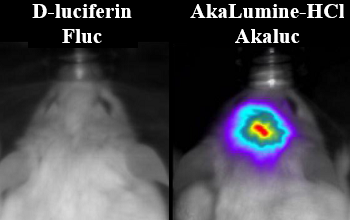
Mice were transfected with the artificial enzymes Fluc and Akaluc into the striatum using Adeno-associated virus (AAV). After 2 weeks, mice were administered intraperitoneally D-luciferin (100 mM) and AkaLumine-HCl (30 mM) and observed head. As a result, mice using AkaBLI which is a combination AkaLumine-HCl and Akaluc showed high luminescent emission.
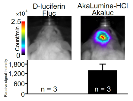
(Fig.5) Top: luminescent images of the head
Bottom: Relative emission intensity (D-luciferin/Fluc or AkaLumine-HCl/Akaluc)
(B) Bioluminescence imaging of Akaluc expression in the striatum of two freely moving mice and marmoset
Mice and marmoset were transfected Akaluc into the striatum. After that, they were administered AkaLumine-HCl at below condition, observed freely moving.
Mice: administered intravenously (75nmol/g)
Marmoset: administered intraperitoneally (75nmol/g)
-
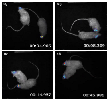
Akaluc expression site: Striatum(Mouse Brain) -
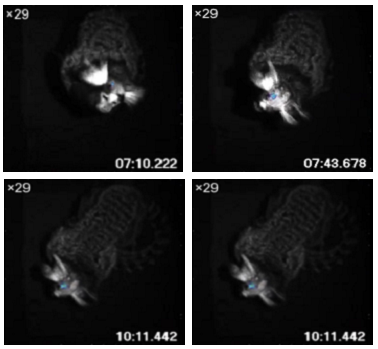
Akalucexpressionsite: Striatum(Marmoset Brain)
Video: Freely moving mice (left) and marmoset (right)
References
- Iwano, S. et al.: Tetrahedron.,69, 3847(2013).
- Kuchimaru,T. et al.: Nature Communications., 7, 11856 (2016)
- Iwano, S. et al.: Science.,359, 935(2018).
Product List
- Open All
- Close All
For research use or further manufacturing use only. Not for use in diagnostic procedures.
Product content may differ from the actual image due to minor specification changes etc.
If the revision of product standards and packaging standards has been made, there is a case where the actual product specifications and images are different.
The prices are list prices in Japan.Please contact your local distributor for your retail price in your region.