See Through Chamber
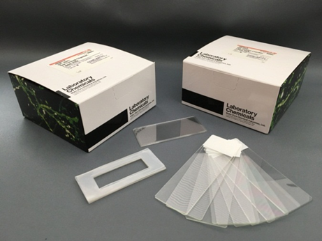
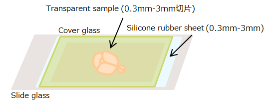
See Through Chamber is a microscopy chamber to be used to assist microscopic observation of optically cleared tissue samples. The product consists of 10 sets of the following: silicone rubber sheet appropriately processed for easy observation of optically cleared samples; cover glass; and slide glass. The silicone rubber sheet is offered with a thickness of 0.3 mm, 0.5 mm, 1.0 mm, 2.0 mm, and 3.0 mm, among which the best one can be selected.
The silicone rubber sheet is covered with protective sheets on both the sides, which can be peeled off before use to facilitate tight contact with slide and cover glasses.
Procedure
Procedure for optical clearing (with SeeDB)
- Fix mouse brain with 4% paraformaldehyde/PBS at 4°C overnight.
- Wash the sample with PBS three times (for 10 minutes each).
- Transfer the sample into a 50-mL conical tube containing 20 mL of SeeDB: 20 w/v% Fructose Solution. Invert the tube on a rotator at room temperature for 4-8 hours (at about 4 rpm). For fragile samples or slices, a seesaw shaker may be used (at about 17 rpm).
- Transfer the sample into a 50-mL conical tube containing SeeDB: 40 w/v% Fructose Solution, and invert it at room temperature for 4-8 hours.
- Transfer the sample into a 50-mL conical tube containing SeeDB: 60 w/v% Fructose Solution, and invert it at room temperature for 4-8 hours.
- Transfer the sample into a 50-mL conical tube containing SeeDB: 80 w/v% Fructose Solution, and invert it at room temperature for 12 hours.
- Transfer the sample into a 50-mL conical tube containing SeeDB: 100 w/v% Fructose Solution, and invert it at room temperature for 12 hours.
- Transfer the sample into a 50-mL conical tube containing SeeDB, and invert it at room temperature for 24 hours.

Figure 1. Optical clearing of biological samples using SeeDB
Examples of samples optically cleared using SeeBD. From left to right, mouse fetus (at embryonic day 12), neonatal mouse whole brain (at postnatal day 3), and adult mouse brain slice (at 8 weeks of age; thickness, 2 mm) are shown.
Mounting
- Select an appropriate See Through Chamber for the actual sample size.
- If SeeBD is used, mount the sample using SeeDB solution.
*Samples which are small and movable during observation will need to be fixed using agarose gel etc.
Observation 1
- Observe SeeDB-treated brain sample using confocal laser microscope or two-photon excitation microscope.
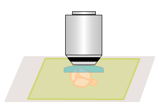
*The refractive index of samples optically cleared with SeeDB is as high as 1.49. Therefore, glycerin-immersion lens, oil- immersion lens, and lens dedicated for optically cleared samples are recommended as the objective lens, but water-immersion lens are also acceptable up to the depth of about 2 mm. Please use the manufacturer’s recommended immersion liquid. Do not use SeeDB solution as the immersion liquid. If images are obtained using dry lens (RI, 1.0) or water-immersion lens (RI, 1.33), correction of depth value will be needed (see reference literature for correction formula).
Observation 2
Examples of sample observation
 |
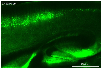 |
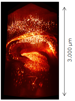
| Figure 2. Fluorescence image of Thy1-YFP-H mouse (10 weeks of age) brain obtained using two-photon excitation microscope | Figure 3. Fluorescence image of Thy1-YFP-H mouse (10 weeks of age) brain obtained using confocal microscope |
References
- Ke, M. T., Fujimoto, S. and Imai, T. : Nat Neurosci ,6 (8),1154 (2013).
- Ke, M. T., Fujimoto, S. and Imai, T.: Bio-protocol 4(3), e1042 (2014).
- Ke, M. T., and Imai, T. : Curr Protoc Neurosci ,66,2.22.1-2.22.19 (2014).
Product List
- Open All
- Close All
For research use or further manufacturing use only. Not for use in diagnostic procedures.
Product content may differ from the actual image due to minor specification changes etc.
If the revision of product standards and packaging standards has been made, there is a case where the actual product specifications and images are different.
The prices are list prices in Japan.Please contact your local distributor for your retail price in your region.


