MagCapture™ Exosome Isolation Kit PS Ver.2
- for Genetic Research
- Manufacturer :
- FUJIFILM Wako Pure Chemical Corporation
- Storage Condition :
- Keep at 2-10 degrees C.
-

 Close
Close
Close
Close -
 Close
Close
Close
Close
- Structural Formula
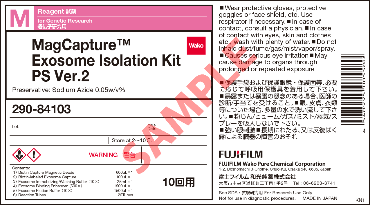
- Label
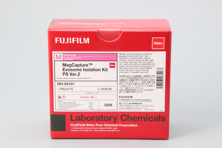
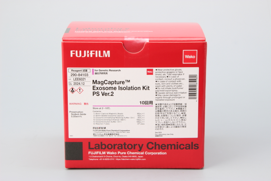
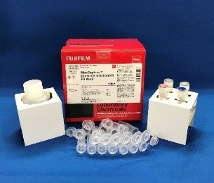
- Packing
|
Comparison
|
Product Number
|
Package Size
|
Price
|
Inventory
|
|
|---|---|---|---|---|---|
|
|
|
2Tests
|
|
In stock in Japan |
|
|
|
|
10Tests
|
|
In stock in Japan |
Document
Kit component
2 tests
| Biotin Capture Magnetic Beads | 120 μL |
|---|---|
| Biotin-labeled Exosome Capture | 20 μL |
| Exosome Immobilizing/Washing Buffer (10x) | 5 mL |
| Exosome Binding Enhancer (500x) | 300 μL |
| Exosome Elution Buffer (10x) | 300 μL |
| Reaction Tubes | 4 tubes |
10 tests
| Biotin Capture Magnetic Beads | 600 μL |
|---|---|
| Biotin-labeled Exosome Capture | 100 μL |
| Exosome Immobilizing/Washing Buffer (10x) | 25 mL |
| Exosome Binding Enhancer (500x) | 1,500 μL |
| Exosome Elution Buffer (10x) | 1,500 μL |
| Reaction Tubes | 22 tubes |
Product Overview
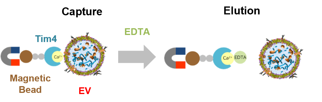
MagCapture™ Exosome Isolation Kit PS Ver.2 is a kit that combines the PS affinity method with magnetic beads for the isolation and purification of extracellular vesicles (EVs), such as exosomes. Purification of intact EVs by elution with a chelating agent was achieved by utilizing Tim4 protein, which binds to phosphatidylserine (PS) on the surface of EVs in a calcium-dependent manner.
Features
- High purity EVs can be obtained with a higher yield than by conventional ultracentrifugation
- Intact EVs can be obtained and used for various applications
- Magnetic beads enable simple operation, achieving multi-sample processing and high reproducibility
Sample
- Cell culture supernatants
- Serum
- Plasma (Heparin, EDTA, Citrate)
- Urine
- Saliva etc.
* Purification of EVs from cerebrospinal fluid has been reported.
Principle
The PS affinity method, Fujifilm Wako’s proprietary EV isolation technique, utilizes the Tim4 protein, which specifically binds to phosphatidylserine (PS) present on the surface of EVs. The high specificity of the PS-Tim4 interaction, coupled with the gentle elution by a chelating agent, enables the isolation of high-purity EVs in their intact state.
About commercial or for-profit purposes
Please use this product for research purposes only. If you wish to use it for commercial or for-profit purposes, please contact Fujifilm Wako at ffwk-labchem-tec@fujifilm.com.
Protocol
The following is a summary of the protocol. For the detailed protocol, please refer to the “Package Insert” on this page.
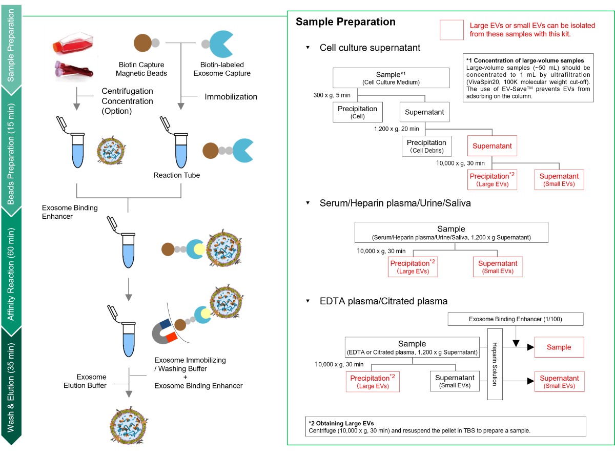
- Outline of Procedure of MagCapture Exosome Isolation Kit PS Ver.2
(Youtube, 7:48)
Data
Comparison with Conventional Methods
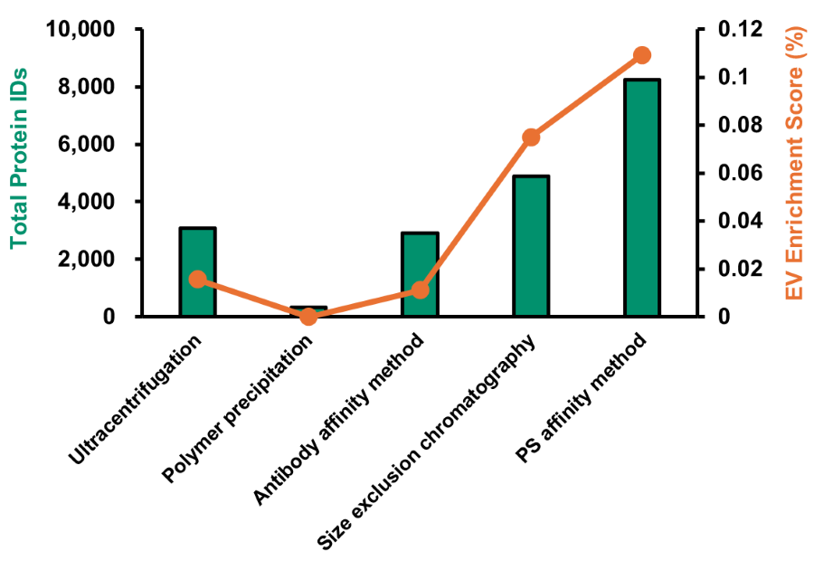
Comparison of the Number of Proteins Identified and EV Enrichment Scores in Human Plasma-Derived EVs by Proteomic Analysis
EVs were isolated from 200 μL of human plasma using each method and subjected to proteomic analysis with an LC/MS Orbitrap Fusion Lumos equipped with FAIMS Pro (DIA, 120 min). EV purity was evaluated by comparing the proportion of identified EV marker proteins relative to the total identified proteins (EES: EV Enrichment Score; see formula below).

[Result]
Compared with other methods, this product yielded a greater number of identified proteins. In addition, the higher EES indicated that EVs of high purity were successfully isolated.
This product is ideal for biomarker discovery using EV-derived proteins in blood samples.
Comparison of Recovery Rates with SEC
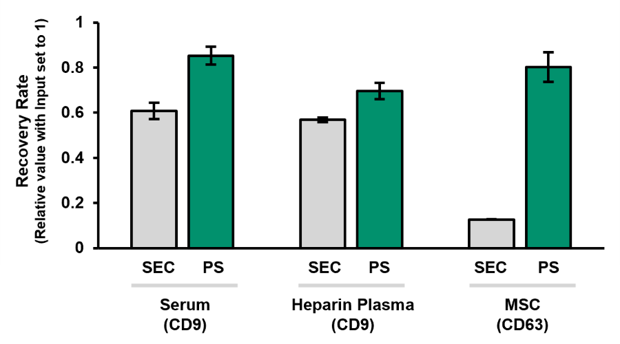
EVs were isolated and purified from human serum, heparin plasma and bone marrow–derived mesenchymal stromal cell/mesenchymal stem cell (MSC) culture supernatants using size exclusion chromatography (SEC) or this product (PS). The EV recovery yield was measured using CD9- or CD63-Capture Human Exosome ELISA Kits (Streptavidin HRP).
[Result]
This product demonstrated a higher EV recovery rate than SEC.
Comparison of anti-inflammatory activity with EVs isolated by ultracentrifugation
IFN-γ-stimulated adipose-derived MSCs (ADMSCs) were cultured in EV production medium EV-Up™ MSC EV Production Basal Medium, AF (Product No. 053-09451) supplemented with additives. EVs were then isolated and purified from the cell culture supernatant using either this product or ultracentrifugation. The isolated EVs were added to mouse primary monocytes, and the expression levels of the inflammatory marker TNF-α (M1 marker) and the anti-inflammatory marker CD206 (M2 marker) were quantified by real-time PCR.
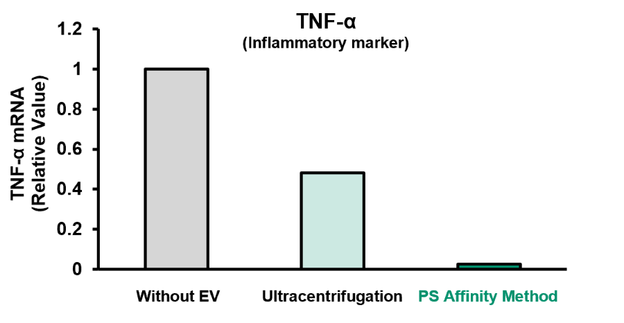
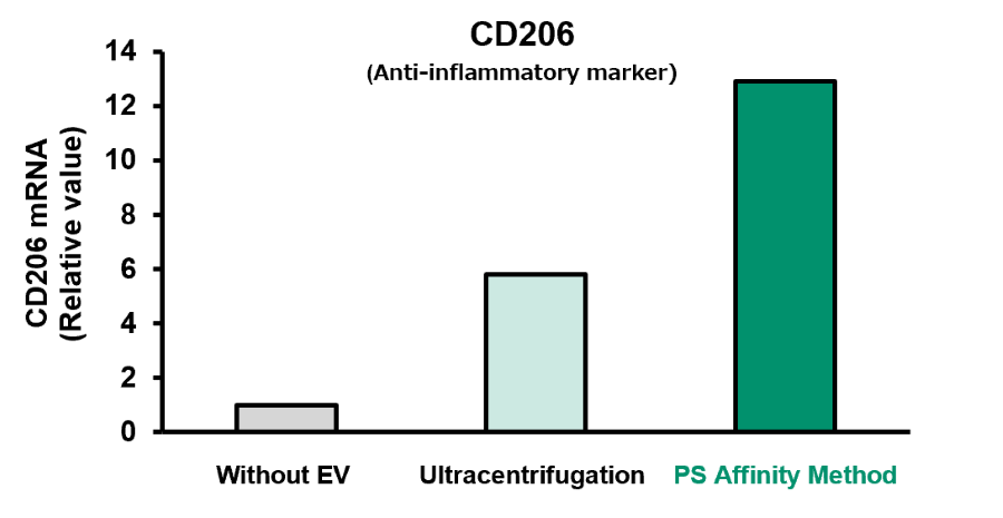
[Result]
EVs obtained using this product (PS affinity method) had higher anti-inflammatory activity than those obtained by ultracentrifugation.
Isolation and Purification of EVs from Various Samples
Serum / Plasma
EVs were isolated and purified from various human blood samples using this product. The EV recovery yield was measured using the PS Capture™ Exosome ELISA Kit (Streptavidin HRP) (Product No. 298-80601). Elution Buffers at two different concentrations (1x and 2x) were evaluated.
[Result]
Using this product, EVs were efficiently recovered from human serum and plasma samples. In particular, recovery from plasma samples was improved when a high-concentration (2x) Elution Buffer was used.

Cell Culture Supernatant
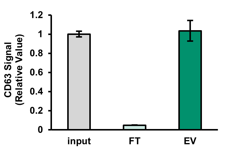
>HEK293
HEK293 cells were expanded in D-MEM containing 10% FBS and then cultured for 3 days in EV production medium EV-Up™ MSC EV Production Basal Medium, AF (Product No. 053-09451) supplemented with additives. EVs were then isolated and purified from the cell culture supernatant using this product. The CD63 signal in the Input, Flow-through (FT), and isolated EV solution (EV) was quantified using the CD63-Capture Human Exosome ELISA Kit (Streptavidin HRP) (Product No. 290-83601), and EV recovery was compared.
[Result]
High EV recovery was achieved with MagCapture™ Exosome Isolation Kit PS Ver.2.
>COLO201
EVs were isolated and purified from COLO201 cell culture supernatant using this product. The isolated EVs or an equal volume of elution buffer (50 μL) was added to pre-seeded normal human fibroblasts, and changes in cell morphology were assessed at 48 hours.
- Purified EV
(0.7x10⁷ particles)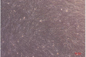
- Elution buffer
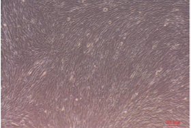
- Untreated
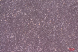
[Result]
No significant changes in cell morphology or notable cell death were observed when EVs isolated and purified with this product or the elution buffer alone was added. These findings suggests that the eluted EVs can be applied directly to cells without additional processing.
Note: The elution buffer contains EDTA. If EDTA may interfere with downstream applications, buffer exchange by ultrafiltration or a similar method is recommended.
References
- Nakai, W. et al.: Sci. Rep., 6(1), 1(2016).
A novel affinity-based method for the isolation of highly purified extracellular vesicles
FAQ
About PS affinity method
- What is the principle of this product?
- This product utilizes Tim4 protein to isolate extracellular vesicles (EVs) including exosomes. Tim4 binds to phosphatidylserine (PS) exposed on the EV membrane surface in a metal-ion-dependent manner.
- What types of extracellular vesicles can be purified?
- This product can be used to purify exosomes and microvesicles with phosphatidylserine exposed on their membrane surface.
- Is it possible to purify exosomes and microvesicles separately?
- Exosomes and microvesicles cannot be completely separated, because they are not clearly distinguishable by size.
The following simple separation method using PS affinity method is recommended to obtain the major fractions for each:
When purifying small extracellular vesicles (small EVs) such as exosomes, use the supernatant after centrifugation at 10,000 x g as the sample.
To purify large extracellular vesicles (large EVs), including microvesicles, first collect the supernatant after centrifugation at 1,200 x g, and then collect the pellet after centrifugation at 10,000 x g. Suspend the pellet in TBS for use as a sample.
If both are purified together, use the supernatant after centrifugation at 1,200 x g as the sample.
- Is anything other than EVs isolated?
- Enveloped viruses are also collected. Since phosphatidylserine is also exposed on the surface of enveloped viruses, both are recovered from samples contaminated with enveloped viruses. Due to this property, PS affinity method may be used to isolate enveloped viruses. The purification of viruses using the PS affinity method was reported in the following paper:
[References]
Santiana, M. et al.: Cell Host & Microbe, 24, 208(2018).
To isolate a virus, affinity purification with an antibody specific to the virus must be performed after collection with PS affinity method. This is true not only for Fujifilm Wako’s kit, but also for purification methods using antibodies against EV markers such as CD63 that is also present on the envelope.
- Do all EVs have exposed phosphatidylserine (PS)?
- Although no finding has been obtained that indicates that all EVs have exposed PS, a comparison of the number of EVs in a sample before and after purification by the PS affinity method using the NTA and ELISA methods shows that about 90% of EVs were recovered from all cell or body fluid samples tested. EVs purified by the PS affinity method showed a higher percentage of low anionic EVs and a lower percentage of high anionic EVs compared to the EVs purified by the ultracentrifugation and size exclusion methods.
- What are the advantages of this method compared to other EV isolation methods?
- Comparison to Ultracentrifugation
High purity EVs can be easily and reproducibly recovered with high efficiency. EVs have been recovered from samples that are not suitable for precipitation by ultracentrifugation. The purity of the recovered EVs is also high; the purity is equivalent to that of the EVs purified by combined ultracentrifugation and density gradient centrifugation methods.
Comparison to Polymer Precipitation
The yield is higher, and high-purity EVs can be obtained compared to polymer precipitation.
Comparison to Antibody Affinity Methods
The antibody affinity method uses antibodies against surface antigens of EVs and requires elution with a denaturing agent or dissociation under acidic conditions for release and recovery of EVs. With PS affinity method, elution is done with a chelating agent under neutral conditions, allowing for recovery of intact EVs. Also, contamination of proteins nonspecifically adsorbed on the beads is low, and high purity EVs can be recovered efficiently.
About Sample
- What types of samples can be used for purification?
- EVs have been recovered from cell culture supernatants, serum, plasma (heparin and EDTA), urine, and feces. Users have also reported purification of EVs from cerebrospinal fluid and saliva. Regarding milk-derived EVs, annexin V present in milk may bind to PS on the EV surface, potentially inhibiting binding to Tim4.
- What animal species are suitable for this kit?
- EVs have been isolated from samples derived from humans, mice, rats, dogs, and monkeys.
- What is the minimum volume of sample needed for purification?
- To ensure consistent mixing of the beads and samples, use at least 500 µL of sample when the reaction is performed using a rotator, or 100 µL when the reaction is performed using a tube mixer. If a smaller volume of sample is used, TBS should be added to bring the volume to the minimum level before reacting with the Exosome Capture-coated beads. It is recommended to add EV-Save™ Extracellular Vesicle Blocking Reagent (Product No. 058-09261) to the TBS used for filling up the samples.
- Is it possible to recover EVs from large-volume samples?
- Approximately 15 mL of cell culture supernatant can be isolated and purified without concentration. Additionally, by performing concentration and other processes, you can accommodate samples of up to 50 mL. Concentrate 50 mL of pre-centrifuged cell culture supernatant by ultrafiltration to 1 mL (recommended filter: Sartorius Vivaspin 20, molecular weight cut-off 100 K, Product No: VS2041). Not only serum-free medium, but medium containing 10% FBS can be used. Serum samples cannot be concentrated, and up to 1 mL can be used.
Fujifilm Wako also offer the MassivEV™ EV Purification Column PS, a column designed for the large-scale purification of EVs from cell culture supernatants using the PS affinity method.
- Molecular weight cut-off of 100 K is recommended for concentration by ultrafiltration, but is it possible to use 10 K?
- Ultrafiltration filters with cut-off values of 100 K, 300 K, and 1,000 K were tested at Fujifilm Wako, and a 100 K filter is recommended based on the concentration time and the amount of EVs concentrated. Filters with smaller cut-off values of 10 K and 30 K could be used, but longer concentration times may be required. For media containing albumin, yield of EVs may be reduced due to concentrated albumin.
- Is there a recommended heparin concentration for plasma (EDTA or citrate) samples?
- Fujifilm Wako adds heparin to achieve a final concentration of 5 mU. Although measurement is possible without heparin addition if the sample is diluted at least five-fold, aggregation may occur during dilution. Therefore, the addition of heparin is recommended.
About Yield
- How many EVs can be isolated from each purification process?
- Although it varies greatly depending on the type and volume of the sample, about 30 μg/mL of protein (measured by the BCA method) and 1-2 x 10¹⁰ particles/mL (measured by NanoSight) in a purification process were obtained when 5 mL of K562 cell culture supernatant (EV secretion was stimulated by monensin sodium salt) was concentrated to 1 mL and used for purification. In addition, about 34 μg/mL of protein and 5 x 10⁹ particles/mL have been recovered from 1 mL of pooled normal human serum in a purification process.
- What is the maximum yield of EVs?
- The maximum yield of one purification process is approximately 1-5 x 10¹⁰ particles.
- Does one Biotin Capture Magnetic Bead bind to only one Biotin-labeled Exosome Capture molecule? Also, how many EVs can be captured by a single Exosome Capture-immobilized bead?
- Multiple Biotin-labeled Exosome Capture molecules bind to a single Biotin Capture Magnetic Bead. Therefore, multiple EVs are captured by a single Exosome Capture-immobilized bead.
About Kit Components and Procedure
- What is the composition of the elution buffer?
- It is a PBS-based solution containing 1 mmol/L of chelating agent and salts. If these ingredients interfere with subsequent analysis, replace the buffer with an appropriate buffer by ultrafiltration (Sartorius VivaSpin500, molecular weight cut-off 100 K, Product No. VS0141) or gel filtration.
- What steps should be performed with particular care?
- In the final part of the washing step after the reaction of the sample with the Exosome Capture-coated beads, the washing solution should be thoroughly removed. Proceed to the elution step after ensuring that the washing solution has been completely removed. In the elution step, suspend the beads thoroughly after adding the elution solution, making sure the beads are not aggregated.
- Is there a step that allows the purification procedure to be carried over to the next day?
- The reaction step with the sample and the Exosome Capture-coated beads can be done overnight.
- Is it possible to reuse the used Exosome Capture-coated magnetic beads?
- It is possible. Used magnetic beads can be reused up to 4 times to recapture the remaining EVs in the sample. The kit is supplied with enough buffer for up to 50 reactions (in the case of a 10-reaction kit) when the same sample is extracted repeatedly, or when there is no concern about cross-contamination. The reuse is recommended for recovery from samples with volumes greater than 1 mL or from concentrated samples.
- Is it possible to store Exosome Capture-coated magnetic beads?
- It is possible. When reusing Exosome Capture-coated beads after elution of EVs, store them in the Washing Buffer supplied with the kit or in TBS prepared separately, and under refrigeration.
About Application
- What types of analysis are possible with the isolated EVs?
- Intact EVs are obtained and can be used for various analyses.
[Example]Protein analysis SDS-PAGE, Western blotting, Proteomics analysis, Flow cytometry, ELISA Genetic analysis qPCR, Microarray, NGS Particle analysis Electron microscopy, Nano-tracking analysis Functional analysis in vitro or in vivo administration
- What amount of EVs is required for each experiment?
- Below are the results of in-house testing.
Experiment Sample amount (example) Electron microscopy 2-4 x 10¹⁰ particles/mL Microarray COLO201: 4.6 x 10¹⁰ particles
TIG-3: 1.7 x 10¹⁰ particles
iPSC: 1.9 x 10⁹ particlesProteomics analysis approximately 1 μg of purified EV Western blotting 15 µL amount out of 100 µL of eluate was used.
- How should purified EVs be stored?
- Add EV-Save™ Extracellular Vesicle Blocking Reagent and store refrigerated or frozen. For long-term storage, store at −80°C. EVs remain stable for 12 months when stored with EV-Save™ Extracellular Vesicle Blocking Reagent. Note that EV-Save™ must be removed prior to proteomic analysis, as it contains a polymer.
- Is it possible to add the isolated EVs directly to the cells in subsequent experiments?
- EVs isolated using this kit have reduced cytotoxicity compared to those using the previous kit, and can be used in in vitro and in vivo experiments. However, if EDTA in the elution buffer causes a problem, the buffer should be replaced.
Troubleshooting
- Purification has been unsuccessful. What can be done?
- Please prepare positive controls. Positive controls can be obtained by culturing any cells such as HEK293, preparing the required amount of cell culture supernatant, and isolating EVs using the kit. Also, the amount of EVs in the medium may be low. Increase scale of the culture.
- How can EV yield be improved?
- For some samples, reducing the amount of immobilized Biotin-labeled Exosome Capture compared to the standard protocol may improve EV yield. For samples with low yield under the standard protocol, try reducing the amount of Biotin-labeled Exosome Capture (e.g., 10 μL → 2-5 μL).
- The total protein content of EV samples purified with this kit is lower than that of other isolation methods. What is the cause of this?
- The total protein content is higher in EV samples recovered by other methods because of the large number of impurities. In contrast, the total protein content of the EV samples purified with this kit is lower, but the actual amount of EVs obtained is not different compared to other isolation methods, because of the high purity of the samples.
- The magnetic beads do not stick to the side of the magnetic stand. Are there any possible causes?
- It is possible that iron, a component of magnetic beads, has been oxidized by a component in the sample that acts as a chelating agent (e.g., citric acid), resulting in decreased adsorption capacity.
Overview / Applications
Property
Manufacturer Information
Alias
For research use or further manufacturing use only. Not for use in diagnostic procedures.
Product content may differ from the actual image due to minor specification changes etc.
If the revision of product standards and packaging standards has been made, there is a case where the actual product specifications and images are different.