PS Capture(TM) Exosome Flow Cytometry Kit
- for Genetic Research
- Manufacturer :
- FUJIFILM Wako Pure Chemical Corporation
- Storage Condition :
- Keep at -20 degrees C.
- Structural Formula
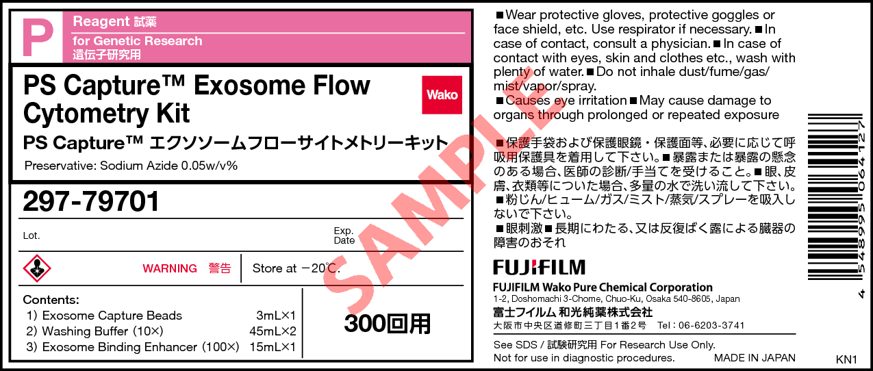
- Label
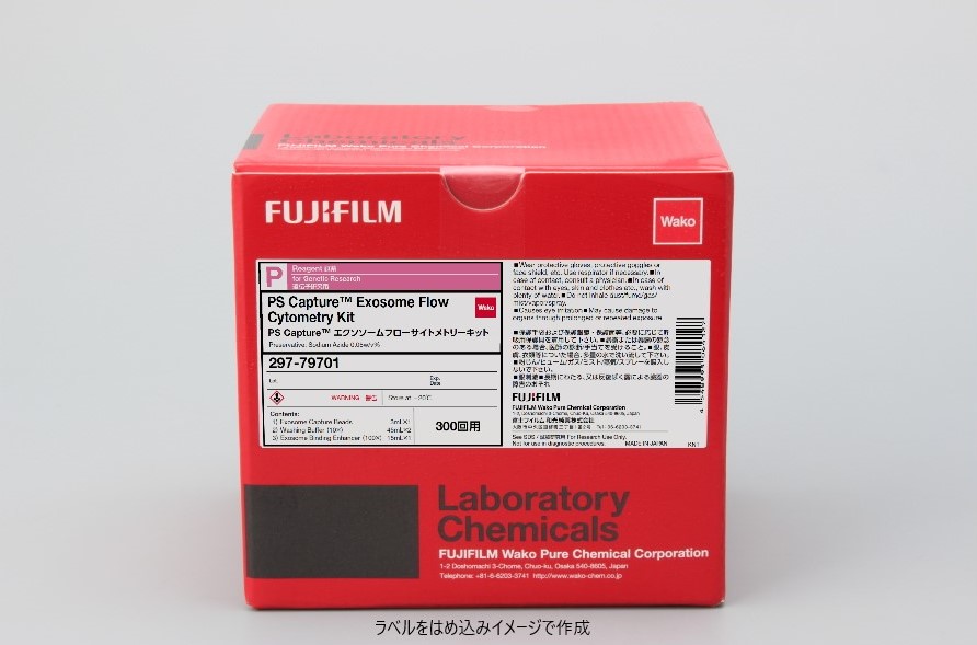
- Packing
|
Comparison
|
Product Number
|
Package Size
|
Price
|
Inventory
|
|
|---|---|---|---|---|---|
|
|
|
300Tests
|
|
In stock |
Document
Kit component
Kit Components (300 assays)
| Exosome Capture Beads | 1 x 3 mL |
|---|---|
| Washing Buffer (10×) | 2 x 45 mL |
| Exosome Binding Enhancer (100×) | 1 x 15 mL |
Overview
Features
- High-Sensitive Qualitative Analysis
- Easy Operation by Magnetic Beads
- Direct Detection without Purification
- Total 3 hours ~ from Isolation to Staining ~
Principle
Procedure
Outline of Procedure ~Basic Protocol for 2 reactions~
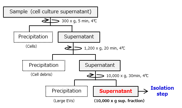
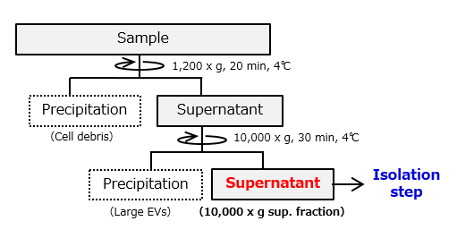
Flowchart of Sample Preparation
-
Cell culture supernatant

-
Serum・Heparin plasma

-
EDTA plasma and Citrated plasma
Pretreatment method of EDTA plasma and Citrated plasma
EDTA and Citric acid, which is the anticoagulant inhibit the binding of extracellular vesicles to Exosome Capture Beads. Therefore, perform pretreatment with sodium heparin solution.
- Prepare 1,000 U/mL sodium heparin solution with purified water. [Example of product used: Code No. 085-00134]
- Add 1 : 200 volume of sodium heparin solution to 100 μL of 10,000 × g supernatant (final concentration : 5 U/mL) and mix.
- Add 1 : 20 volume of Exosome Binding Enhancer (100×) further and mix.
- Proceed to “Isolation step”.
Recommended reaction scale
Basic protocol is set as 2 reactions using a 1.5 mL microcentrifuge tube to isolate EVs from samples with Exosome Capture Beads.
For scale-up, increase the amount of Exosome Capture Beads and samples. Recommended reaction scale is presented in the right table.
Note: 10 reactions are maximum for one 1.5 mL microcentrifuge tube.
| Qty of Reaction | Exosome Capture Beads (µL) | Sample Volume(µL) |
|---|---|---|
| 2 reactions (basic) |
30 | 100 |
| 3 reactions | 40 | 133 |
| 4 reactions | 50 | 167 |
| 5 reactions | 60 | 200 |
| 6 reactions | 70 | 233 |
| 7 reactions | 80 | 267 |
| 8 reactions | 90 | 300 |
| 9 reactions | 100 | 333 |
| 10 reactions | 110 | 367 |
Applications
Qualitative analysis of exosomes in cell culture supernatant of K562 cell line
Exosomes in cell culture supernatant of K562 cells were isolated using PS Capture™ Exosome Flow Cytometry Kit or each of anti-CD81-, CD9- and CD63-antibody-immobilized magnetic beads (supplier A), followed by flow cytometric analysis of exosome surface antigens after immunostaining with fluorescence-labeled antibodies.
| Sample | Cell culture supernatant of K562 cells: 33 μL/Assay |
|---|---|
| Detection antibody | PE-anti-CD63 (BD Biosciences, 556020) PE-anti-CD9 (Novus Biologicals, NB100-77915PE) PE-anti-CD81 (Novus Biologicals, NBP1-44861PE) |
① PS Capture™ Exosome Flow Cytometry Kit
② Anti-CD81 antibody immobilized magnetic beads
③ Anti-CD9 antibody immobilized magnetic beads
④ Anti-CD63 antibody immobilized magnetic beads
Each signal value was normalized using the value of Isotype.
It was confirmed that the PS Capture™ Exosome FCM Kit can detect the exosome surface antigen with high sensitivity compared with competitors' products, regardless of which detection antibody is used.
Qualitative analysis of exosomes in serum and plasma
Exosomes in human serum and human plasma (EDTA plasma, heparin plasma) were isolated using PS Capture™ Exosome Flow Cytometry Kit, followed by flow cytometric analysis of exosome surface antigens after immunostaining with PE-labeled mouse IgG isotype control and PE-labeled anti-human CD9 antibody.
| Sample: 33 μL/Assay each |
Human serum Human heparin plasma Human EDTA plasma (buffer exchanged) |
|---|---|
| Detection antibody | PE-labeled anti-CD9 antibody (Novus Biologicals, NB100-77915PE) |
In any of the samples, the peak shift of fluorescence intensity was confirmed when stained with PE-labeled anti-human CD9 antibody.
Overview / Applications
| Outline | This kit is a reagent that can qualitatively analyze extracellular vesicles contained in cell culture supernatants and body fluid samples by flow cytometry. After extracellular vesicles are reacted and immobilized on magnetic beads on which extracellular vesicle surface phosphatidylserine (PS) specifically binding protein is immobilized, fluorescent labeling for any extracellular vesicle marker protein by using antibodies, we can detect marker proteins on extracellular vesicle surface with high sensitivity. Before using it, prepare the primary antibody for each surface marker protein, the fluorescent dye-labeled secondary antibody, or the primary antibody labeled with fluorescent dye. |
|---|
Property
Manufacturer Information
Alias
For research use or further manufacturing use only. Not for use in diagnostic procedures.
Product content may differ from the actual image due to minor specification changes etc.
If the revision of product standards and packaging standards has been made, there is a case where the actual product specifications and images are different.


