Luminescent Probes
A luminescent probe is a substance whose substrate moves to an excited state through chemical reactions and then returns to a ground state, emitting light. Thanks to the use of chemical reactions, luminescent probe is characterized by a high selectivity, which contributes to low background noise and increases signal detection. Since a luminescent probe covers a wide dynamic range, it is often used to investigate the dynamics of ATP and calcium ions. It is also used as a reporter gene to visualize specific cells or investigate gene expression. Fujifilm Wako sells AkaLumine-HCl and coelenterazine, which perform better than the substrates used in conventional luminescent probes.
Product Line-up
More Information
Types and Features of Luminescent Probes
Firefly Luciferase/FLuc
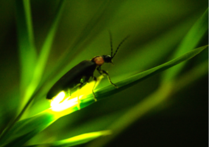
Firefly luciferase is a bioluminescent enzyme that uses luciferin as a substrate. Luciferase converts luciferin to adenylyl luciferin in the presence of ATP and Mg2+. Adenylyl luciferin reacts with O2 and, via an intermediate, emits light at a wavelength of about 560 nm.
Firefly luciferase from North America emits a yellow-green light (560 nm) in neutral to slightly alkaline solutions and a weak red light (615 nm) in acidic solutions. Therefore, a red light is emitted in acidic organelles at greatly reduced intensity. Luciferases cloned from click beetles and railroad worm emit high intensity light even under acidic conditions.
Firefly luciferase is a commonly used bioluminescent enzyme, but the emitted light at 560 nm (i.e. its luminescence peak) is prone absorption by living tissue, making deep tissue imaging difficult. Fujifilm Wako offers AkaLumine-HCl, an artificial substrate alternative to luciferin. AkaLumine-HCl emits light in the near-infrared region (670-680 nm), which is less susceptible to absorption by water and hemoglobin, and thus allows for deeper tissue imaging.
Aequorin
Aequorin is a luminescent protein derived from the jellyfish Aequorea aequorea. It is composed of the protein portion apoaequorin, the substrate coelenterazine, and molecular oxygen O2.
The luminescence of aequorin is calcium ion-dependent and is used to measure intracellular calcium ions. When a calcium ion binds to aequorin, coelenterazine is oxidatively decomposed, producing excited carbonyl. When the excited carbonyl returns to the ground state, the energy is emitted as light (about 465 nm).
Aequorin emits green light by bioluminescence resonance energy transfer (BRET) when GFP is present in close proximity. This mechanism is used to analyze protein-protein interactions.
Fujifilm Wako offers coelenterazine h, a synthetic coelenterazine derivative. Compared to coelenterazine, coelenterazine h/aequorin complex has 10 times stronger luminescence intensity, lower toxicity, and is highly permeable to the plasma membrane.
Renilla Luciferase/RLuc
Renilla luciferase is a luciferase from the sea pansy (Renilla reniformis). Like aequorin, it uses coelenterazine as a substrate, and its luminescence peaks at about 480 nm. Since the substrate of renilla luciferase is different from the substrate of firefly luciferase, these enzymes are used in dual reporter assays, in which two detection systems are used simultaneously.
Cypridina Luciferase/CLuc
Sea firefly luciferase is a bioluminescent enzyme from the sea firefly (Cypridina noctiluca). It uses sea firefly luciferin as a substrate, and its luminescence peaks at about 460 nm. Sea firefly luciferase is a secreted protein and can be obtained without cell disruption. It is also more resistant to detergents such as sodium dodecyl sulfate than copepod luciferase (see below).
Gaussia Luciferase/GLuc
Gaussia luciferase is a bioluminescent enzyme derived from the crustacean Copepods (genus Gaussia), a well-known zooplankton. It uses coelenterazine as a substrate, and its luminescence peaks at about 480 nm. Like sea firefly luciferase, it is a secreted protein and can be used for high-throughput screening.
References
- "How to select and use fluorescent and luminescent reagents for successful experiments” ed. by Miwa, Y., Yodosha, Japan, (2007). (Japanese).
- Akimoto, H. et al.: Seibutsu Butsuri, 49(2), 070(2009).
Bio-Photonic Imaging using a Bioluminescence System (Japanese) - "Life Science Reagent Utilization Handbook” ed. by Tamura, T., Yodosha, Japan, (2009). (Japanese).
For research use or further manufacturing use only. Not for use in diagnostic procedures.
Product content may differ from the actual image due to minor specification changes etc.
If the revision of product standards and packaging standards has been made, there is a case where the actual product specifications and images are different.