Glycobiology
Glycans in living organisms are mainly linked to proteins and lipids, and contribute to their various biological functions. The importance of glycans has been reported in various fields such as cancer, immunology, development/differentiation, regenerative medicine, and infectious diseases. Fujifilm Wako offers various lectins, anti glycan antibodies, and other tools for glycan research and analysis.
More Information
What Are Glycans?
Glycans are chains of sugars linked by glycosidic bonds and are called the "third chain" after nucleic acids and proteins. Glycans form much more diverse structures than nucleic acids and proteins, due to their large number of linkage sites.
Glycans in living organisms are mainly linked to proteins and lipids, and in this state are called glycoproteins and glycolipids, respectively. The properties of these molecules are described below.
Glycoproteins
Many proteins are glycoproteins with attached glycans. In particular, most membrane proteins and serum proteins are glycoproteins. Proteins with one or more long polysaccharide chains (glycosaminoglycans) are called proteoglycans.
Glycans of glycoproteins are linked to proteins mainly by two types of linkage, N-linked and O-linked. In the N-linked (Asn type) glycosylation, N-acetylglucosamine (GlcNAc) is linked to the Asn residue in the Asn-X-Ser/Thr sequence by an N-β-glycosidic bond. In addition to GlcNAc, N-linked glycans contain galactose, fucose, mannose, sialic acid, etc., while GalNAc, which is found in the O-linked glycans described below, is rarely present. On the other hand, in the O-linked glycosylation (mucin type), N-acetylgalactosamine (GalNAc) is linked to Ser or Thr by an O-α-glycosidic bond. O-linked glycans contain galactose, fucose, sialic acid, etc. in addition to GalNAc, while mannose, the component N-linked glycans, is usually not present.
Glycans of glycoproteins influence the physical properties of proteins, including solubility, charge, stability, viscosity, higher-order structure, anti-freezing, degradation resistance, and masking of antigenicity.
Glycolipids
Many lipids, like proteins, in the eukaryotic plasma membrane are glycolipids with linked glycans. Glycolipids are broadly classified into sphingoglycolipids and glyceroglycolipids. Sphingoglycolipids are composed of glycans attached to ceramide (a lipid consisting of sphingosine and a fatty acid), while glyceroglycolipids are composed of glycans attached to 1,2-diacylglycerol (glycerol backbone with linked fatty acids).
Glycolipids in the plasma membrane cover the cell surface together with glycoproteins and proteoglycans, described above. Glycans on the cell surface differ depending on the cell type and are used for cell identification. Glycolipids themselves have various activities; they are known to induce cell differentiation and regulate cell proliferation by controlling activities of proteins.
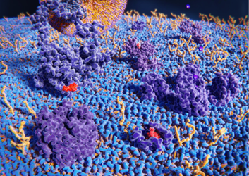
Glycans Are the “Face of the Cells”
Eukaryotic cells are covered with glycans. Because the composition of glycans varies depending on the type and nature of the cells, they are called the “face of the cells.”
In fact, organisms use glycans to recognize and identify cells. For example, sperm cells use glycoproteins present on the surface of the egg to distinguish between oocytes and non-oocytes. When infected with pathogens, endothelial cells express lectins on the surface of the plasma membrane to identify the surface glycans of neutrophils, and selected neutrophils then migrate to the site of infection. Furthermore, the structure of cell surface glycans differs greatly between differentiated cells (somatic cells) and undifferentiated cells (ES cells and iPS cells).
There are also individual differences among glycans, the best-known example being blood types. The ABO blood group is defined by differences in the structure of glycans on the surface of red blood cells. When organ transplants are performed between humans with different blood types, rejection is triggered because the cells have different faces.
Glycans on the cell surface are also closely associated with pathogens and cancer. For example, hemagglutinin, which is present on the membrane surface of influenza viruses, binds to glycans (sialic acid) on the cell surface and enters the cell. The glycan structure of cancer cells differs from that of normal cells. Specific glycans that increase during carcinogenesis are known to enhance the metastatic potential of cancer.
The use of glycans as the face of the cells makes it possible to detect and remove specific cells, and anti-glycan antibodies and lectins that recognize specific glycans are being developed.
References
- Glycoengineering Editorial Board: “Glycoengineering”, Industrial research committee, (1992). (Japanese)
- Alberts, B., et al: “Essential Cell Biology 2nd ed.”, Garland Science/Taylor & Francis Group, (2003).
- Narimatsu, H.: Synthesiology, 5(3), 190(2012).
Development of basic tools for glycoscience and their application to cancer diagnosis
For research use or further manufacturing use only. Not for use in diagnostic procedures.
Product content may differ from the actual image due to minor specification changes etc.
If the revision of product standards and packaging standards has been made, there is a case where the actual product specifications and images are different.