



Anti c-Fos Antibodies
c-Fos gene is well-known as an "immediate early gene" because it is among the first genes to be induced in response to cellular stimulation. In neuroscience, c-Fos is widely used as a marker of neuronal activity. Although the target genes regulated by c-Fos in neurons have not yet been fully characterized, c-Fos is thought to be involved in regulating the expression of many neuropeptide and neurotransmitter-synthesizing enzyme genes.
Fujifilm Wako offers a polyclonal antibody against c-Fos raised in guinea pigs. It can be used to detect neuronal activity by immunohistochemistry. This guinea pig-derived antibody is compatible with multiplex staining alongside rabbit- or mouse-derived antibodies.
What is c-Fos?
c-Fos is a member of the Fos protein family and functions as a transcription factor. Localized primarily in the nucleus, it forms heterodimers with partner proteins such as c-Jun. These complexes bind to promoter regions and regulate the expression of target genes. The c-Fos gene is well-known as an “immediate early gene” because it is among the first genes to be induced in response to cellular stimulation. It is thought to play a key role in initiating stimulus-dependent changes to gene expression.
In neuroscience, c-Fos is widely used as a marker of neuronal activity. Previous studies have shown that c-Fos expression is rapidly induced following neuronal stimulation, with mRNA levels peaking within about 30 minutes. Protein levels subsequently peak 60–90 minutes after stimulation and remain elevated for roughly 2–5 hours1). Although the target genes regulated by c-Fos in neurons have not yet been fully characterized, c-Fos/Jun binding sites are found in the promoters of many neuropeptide and neurotransmitter-synthesizing enzyme genes, implicating c-Fos in the control of their expression.
Anti c-Fos, Guinea Pig
“Anti c-Fos, Guinea Pig” is a polyclonal antibody against c-Fos raised in guinea pigs. It can be used to detect neuronal activity by immunohistochemistry. This guinea pig-derived antibody is compatible with multiplex staining alongside rabbit- or mouse-derived antibodies.
Antibody Information
| Clonality | Polyclonal |
|---|---|
| Antigen | Synthetic peptide (N-terminus of c-Fos) |
| Host | Guinea Pig |
| Formulation | PBS, 1% Trehalose, 0.05% Sodium azide |
| Conjugate | Unconjugated |
| Cross-reactivity | Mouse |
| Application | Immunohistochemistry (Frozen section) 1:500-2,000 |
Application Data
Immunohistochemistry
>Comparison of c-Fos expression after lipopolysaccharide (LPS) administration
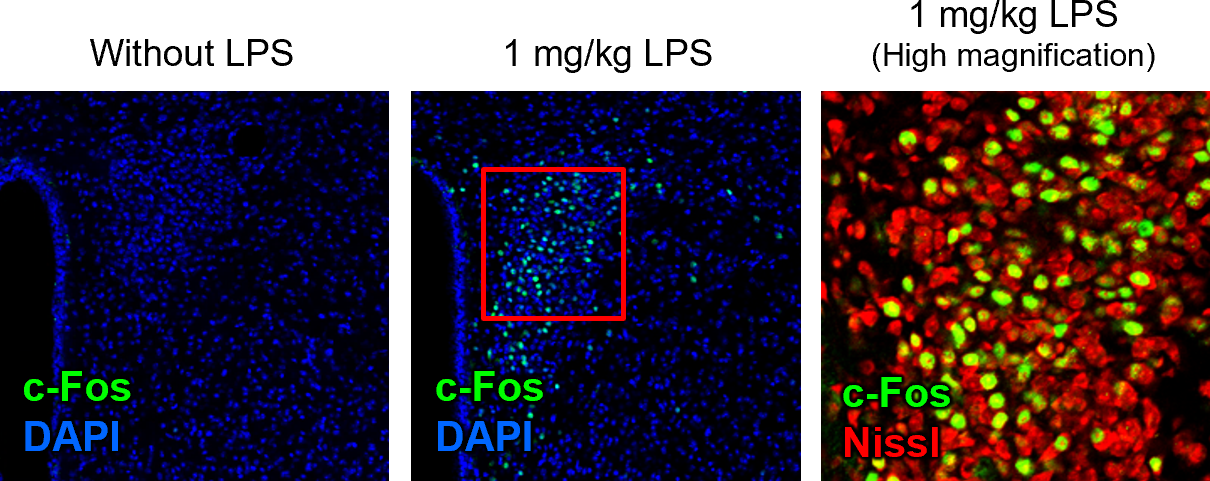
Specie: Mouse
Site: Paraventricular hypothalamic nucleus
Sample: Frozen section
Antibody concentration: 1:1,000
Note: Tissue collected 2 hours after intraperitoneal administration of 1 mg/kg LPS
Data by courtesy of
Dr. Furube, Department of Functional Anatomy and Neuroscience, Asahikawa Medical University
[Result]
No c-Fos-positive cells were observed without LPS stimulation, whereas numerous c-Fos-positive cells appeared 2 hours after LPS administration. Because much of the c-Fos signal colocalized with Nissl-positive cell bodies, c-Fos was confirmed to be expressed in neurons.
>Time course of c-Fos expression under osmotic stimulation
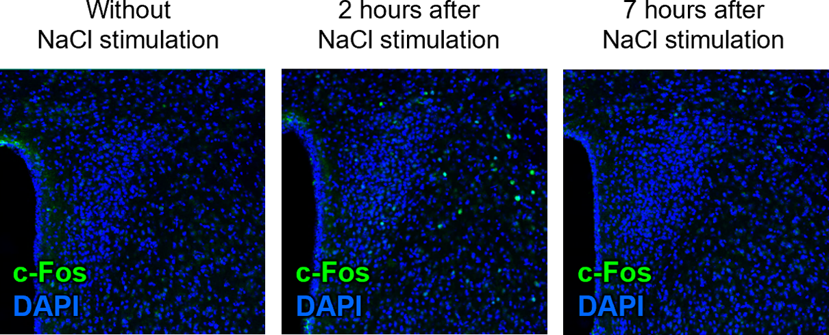
Species:Mouse
Site: Paraventricular hypothalamic nucleus
Sample: Frozen section
Antibody concentration: 1:1,000
Note: Tissue collected 2 hours after intraperitoneal administration of 0.5 mL of 4.5% NaCl.
Data by courtesy of
Dr. Furube, Department of Functional Anatomy and Neuroscience, Asahikawa Medical University
[Result]
Numerous c-Fos-positive cells were observed 2 hours after NaCl stimulation, whereas almost none remained positive at 7 hours—confirming the brief, transient expression pattern characteristic of c-Fos.
>Comparison with Competitors’ Products
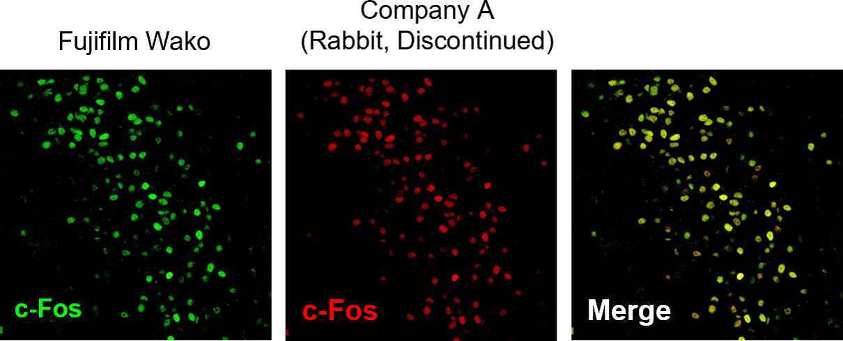
Species: Mouse
Site: Paraventricular hypothalamic nucleus
Sample: Frozen section
Antibody concentration: 1:1,000
Note: Tissue collected 2 hours after intraperitoneal administration of 1 mg/kg LPS
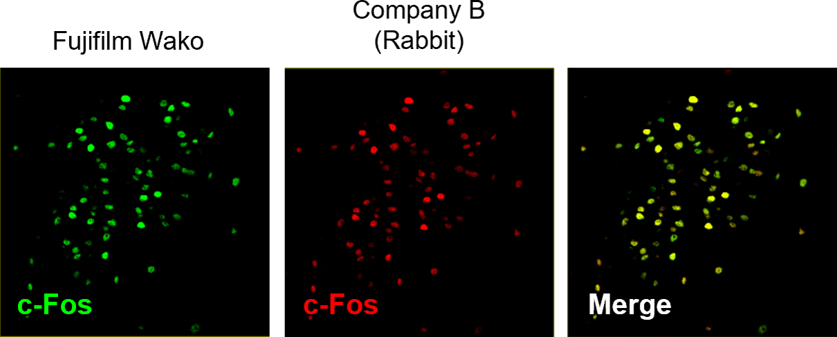
Species: Mouse
Site: Paraventricular hypothalamic nucleus
Sample: Frozen section
Antibody concentration: 1:1,000
Note: Tissue collected 2 hours after intraperitoneal administration of 1 mg/kg LPS
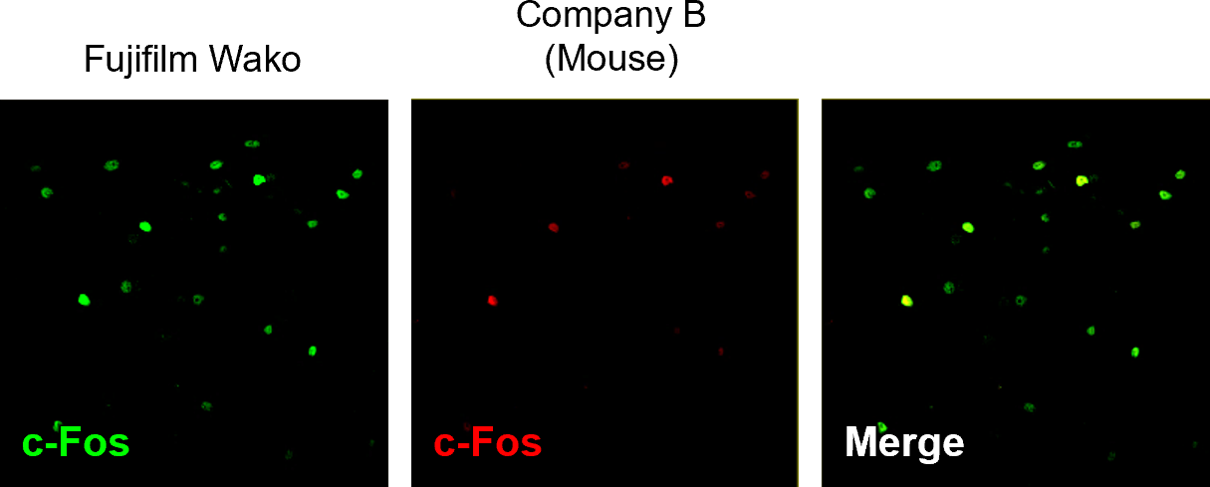
Species: Mouse
Site: Paraventricular hypothalamic nucleus
Sample: Frozen section
Antibody concentration: 1:1,000
Note: Tissue collected 2 hours after intraperitoneal administration of 1 mg/kg LPS
Data by courtesy of Dr. Furube, Department of Functional Anatomy and Neuroscience, Asahikawa Medical University
[Result]
This antibody produced a detection pattern comparable to those of competitor’s rabbit-derived antibodies. It demonstrated higher detection sensitivity than competitor’s mouse-derived antibody.
References
- Hoffman, G. E. et al.: Front. Neuroendocrinol., 14(3), 173(1993).
c-Fos and related immediate early gene products as markers of activity in neuroendocrine systems
Product List
- Open All
- Close All
Anti c-Fos, Guinea Pig (polyclonal antibody)
For research use or further manufacturing use only. Not for use in diagnostic procedures.
Product content may differ from the actual image due to minor specification changes etc.
If the revision of product standards and packaging standards has been made, there is a case where the actual product specifications and images are different.