



Fibronectin, Human, recombinant
Fibronectin, a major component of the extracellular matrix, regulates cell adhesion, migration, proliferation, and differentiation. In cell culture, it serves as an essential matrix for cellular attachment and growth.
We offer recombinant plasma fibronectin and cellular fibronectin produced using genetically modified silkworms. These proteins are derived from cocoons in a human-pathogen-free environment, ensuring high safety.
Conventional Challenges
Plasma Fibronectin
Present in high concentrations in blood, plasma fibronectin plays a key role in blood clotting and wound healing. While blood-derived products have been widely used as cell culture coatings, the use of human or animal blood as a raw material raises concerns regarding potential pathogen contamination.
Cellular Fibronectin
Synthesized by cells in their immediate microenvironment, cellular fibronectin promotes cell adhesion and proliferation; however, obtaining it in a highly purified form has traditionally been difficult.
High-purity purification was achieved using liquid chromatography with an agarose-based matrix free of animal-derived components.
What is Fibronectin?
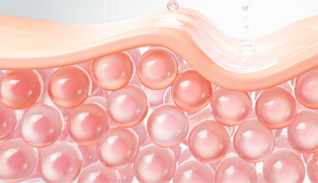
Fibronectin is a glycoprotein widely distributed throughout the extracellular matrix (ECM). It is involved in various biological processes, including cell adhesion, proliferation, differentiation, and migration. Composed of a dimer of approximately 220 kDa subunits, it specifically binds to diverse molecules—such as integrin receptors on the cell surface and ECM components including collagen, fibrin, complement proteins, glycosaminoglycans, and heparin—via its multiple functional domains.
In vivo, fibronectin is produced by mesenchymal cells such as fibroblasts, chondrocytes, macrophages, and hepatocytes, playing a crucial role in maintaining tissue architecture and promoting injury repair. Particularly at wound sites, fibronectin rapidly deposits to form cell attachment sites, which facilitates the migration and proliferation of fibroblasts and vascular endothelial cells to accelerate tissue repair. Additionally, it is stored in platelet alpha-granules and expressed on the platelet surface upon activation, contributing to the hemostatic mechanism.
In the fields of tissue engineering and regenerative medicine, fibronectin is utilized as a matrix for cell culture. Biomaterials containing fibronectin enhance cell attachment and promote 3D tissue formation, supporting biocompatibility and functional tissue reconstruction. As a result, it is expected to improve cell culture efficiency and post-transplantation engraftment rates, leading to various advanced applications.
Features of Fibronectin, Human, recombinant, Plasma / Cellular
- Purified using a high-purity, agarose-based matrix free of animal-derived substances
- Exhibits cell adhesion activity equivalent to or higher than native fibronectin
- Eliminates the risk of pathogen contamination as it is not derived from blood
Product Overview
- Lyophilized
- Bacterial endotoxin tested
- Protein concentration: 450–550 μg/mL
- Sterility tested
- Mycoplasma tested
【Modular Structure】
Fibronectin, Human, recombinant, Plasma
Expressed as a homodimer of a short variant lacking EDA and EDB, but containing the ⅢCS segment.
• Molecular Weight: 499 kDa
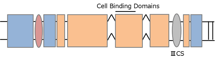
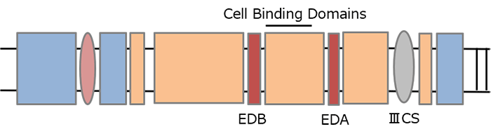
Fibronectin, Human, recombinant, Cellular
Reproduces diverse splicing variants containing EDA, EDB, and ⅢCS segments.
• Molecular Weight: 539 kDa
How to Use
Preparation
Add 2 mL of sterile purified water to the vial and let it stand at room temperature for 30 minutes.
(The resulting fibronectin concentration after reconstitution is 0.5 mg/mL.)
Storage
For storage at -20℃ or lower, aliquot into smaller portions to avoid repeated freeze-thaw cycles.
Stable for approximately 6 months under proper storage conditions.
Coating Protocol (Example)
- Dilute the prepared solution with PBS(-) or a similar buffer, and coat the plate at a concentration of 1-5 µg/cm2.
Example: To coat a 6-well plate (9.6 cm2/well) at 2.5 µg/cm2 (24 µg/well), add 2 mL of fibronectin solution
diluted to 12 µg/mL. - Incubate at 37℃ for 1 hour. Remove the solution, wash with PBS(-), and seed the cells.
Applications
Ideal as a matrix for a wide variety of cultured cells, including mesenchymal stem cells (MSCs). With high safety and minimal lot-to-lot variability in performance, it is well-suited for a broad range of applications, including regenerative medicine research.
Data
Human Bone Marrow-Derived MSCs
Cell Adhesion Assay
Media: Serum-free media for MSC
Fibronectin: 2.5 μg/cm2 each
Seeding Density: 1.58 x 10³ cells/cm2
Culture Conditions: 37℃、5% CO2
Incubation Time: 1 hour
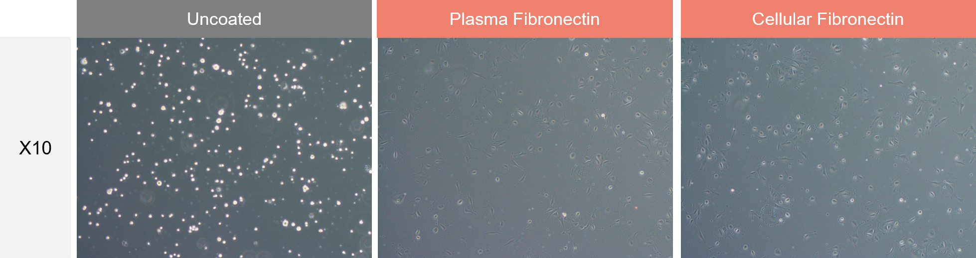
Our fibronectins (plasma fibronectin and cellular fibronectin) were confirmed to promote the adhesion of human bone marrow-derived MSCs to culture vessels.
Cell Proliferation Assay
Media: Serum-free media for MSC
Fibronectin: 2.5 μg/cm2 each
Seeding Density: 5×10³ cells/cm2
Culture Conditions: 37℃、5% CO2
Incubation Time: 3 days
Coating culture vessels with our fibronectins (plasma fibronectin and cellular fibronectin) improved the proliferation of human bone marrow-derived MSCs.
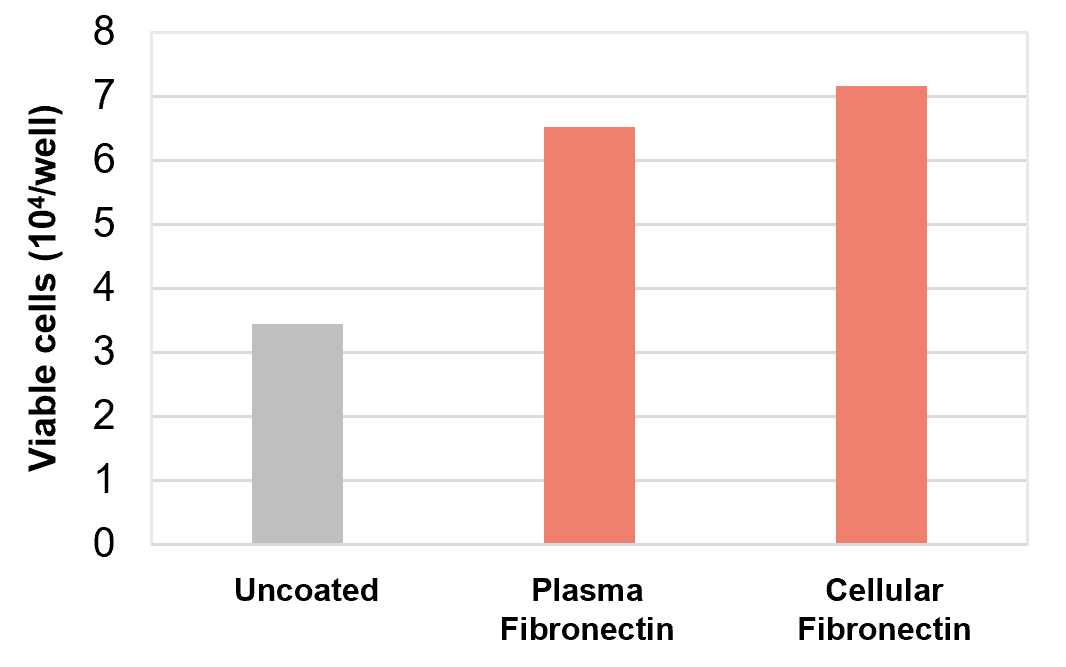
BHK21 Cells
Cell Adhesion Assay
Media: Serum-free medium
Fibronectin: 2.5 μg/cm2 each
Seeding Density: 7.4×10⁴ cells/cm2
Culture Conditions: 37℃, 5% CO2
Incubation Time: 40 minutes
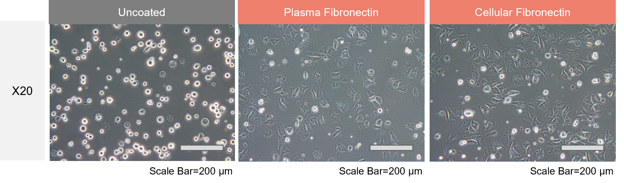
Our fibronectins (plasma fibronectin and cellular fibronectin) were confirmed to promote the adhesion of BHK21 cells to culture vessels.
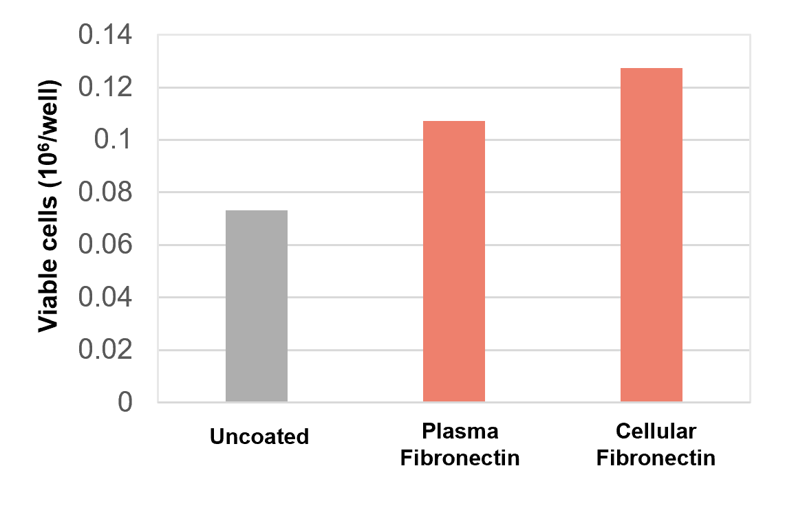
Cell Proliferation Assay
Media: Serum-free medium
Fibronectin: 2.5 μg/cm2 each
Seeding Density: 1×10⁴ cells/cm2
Culture Conditions: 37℃, 5% CO2
Incubation Time: 4 days
Coating culture vessels with our fibronectins (plasma fibronectin and cellular fibronectin) improved the proliferation of BHK21 cells.
Product List
- Open All
- Close All
For research use or further manufacturing use only. Not for use in diagnostic procedures.
Product content may differ from the actual image due to minor specification changes etc.
If the revision of product standards and packaging standards has been made, there is a case where the actual product specifications and images are different.