SCALEVIEW-S4
SCALEVIEW-S creates transparent brains, organs,and cultured cells with minimal tissue damage, that can handle both florescent and immunohistochemical labeling techniques, and is even effective in older animals.
The new technique creates transparent brain samples that can be stored in SCALEVIEW-S solution for more than a year without damage.
Features
- Easy-to-use
- No special equipment required
- Less damage to sample
- Compatible with IF, FP and other fluorescent labels
T47D cell spheroid observation example
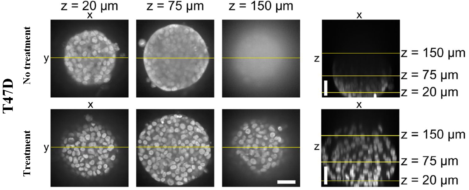
Figure 1. Confocal imaging of T47D cell spheroids after clearing. (nuclei:Hoechst 33342)
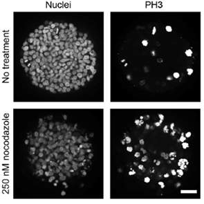
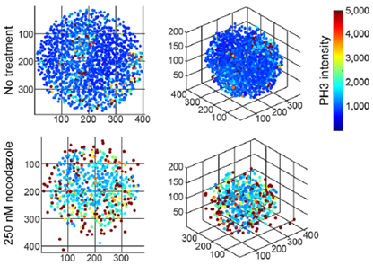
Figure 2. PH3 immunostaining, clearing, and segmentation analysis in nocodazole-treated T47D spheroids.
Data by Molly E, Boutin, et al. : Scientific Reports, 8, 11135 (2018).
Protocol

Application
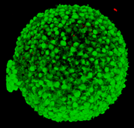
Primary Neurosphere from Adult Rat Hippocampus (5 days in vitro)
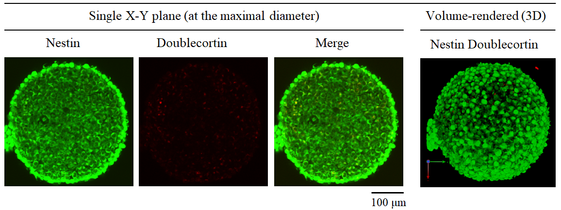
Figure 3. 3D visualization of Neurosphere
Microscope (CLSM) :Olympus FV1000
Objective lens :UMPLFLN10XW (NA 0.3)
References
- Hama,H.et al. : Nature Neuroscience, 14, 1481(2011).
- Hama,H.et al. : Nature Neuroscience, 18, 1518(2015).
- Hama H, et al. : Protocol Exchange (2016), doi:10.1038/protex.2016.019
- Molly E, Boutin, et al. : Scientific Reports, 8, 11135 (2018).
Product List
- Open All
- Close All
For research use or further manufacturing use only. Not for use in diagnostic procedures.
Product content may differ from the actual image due to minor specification changes etc.
If the revision of product standards and packaging standards has been made, there is a case where the actual product specifications and images are different.


