Anti P2RY12, Guinea Pig
- for Immunochemistry
- Manufacturer :
- FUJIFILM Wako Pure Chemical Corporation
- Storage Condition :
- Keep at -20 degrees C.
- Structural Formula
- Label
- Packing
- SDS
|
Comparison
|
Product Number
|
Package Size
|
Price
|
Inventory
|
|
|---|---|---|---|---|---|
|
|
|
10uL
|
|
In stock in Japan |
|
|
|
|
100uL
|
|
In stock in Japan |
Please check here for notes on products and prices.
Document
Product Overview
P2RY12 (also known as P2Y12) is an adenosine 5‘-diphosphate (ADP) receptor and a member of the P2Y family of purinergic G protein-coupled receptors. In the central nervous system, it is specifically expressed in microglia1) but not macrophage, and is used as a homeostatic microglial marker.
The “Anti P2RY12, Guinea Pig” is a guinea pig polyclonal antibody, raised against P2RY12. It can be used to perform multiplex immunohistochemistry.
Antibody Information
| Clonality | Polyclonal |
|---|---|
| Antigen | Synthetic peptide (P2RY12 C-terminal sequence) |
| Host | Guinea pig |
| Formulation | PBS, 0.05% Sodium azide |
| Conjugate | Unconjugated |
| Cross-reactivity | Mouse* |
| Application | Immunohistochemistry (frozen section) 1:500-2,000 |
* Experience with rats has been reported. For details, please see “Data”.
Protocol (Example)
- 1.Perfusion fixation
- Perfuse with sodium citrate/PBS to remove blood, followed by perfusion with 4% PFA.
- 2.Post-fixation
- 4% PFA (-24 hours)
- 3.Immersion in sucrose
- 30% Sucrose/PBS (4℃, overnight - approximately 2 days)
- 4.Preparation of frozen sections
- After removing sucrose, prepare frozen blocks and cut 30-μm thick sections using a cryostat.
- 5.Blocking
- 5% normal goat serum in 0.3% Triton X-100/PBS (4℃, overnight)
- 6.Primary antibody reaction
- Anti P2RY12, Guinea pig (1:1,000), 1% normal goat serum in 0.3% Triton X-100/PBS (4℃, overnight - 2 days).
- 7.Washing
- 0.3% Triton X-100/PBS (5 minutes x 3)
- 8.Secondary antibody reaction
- Alexa Fluor® 488 AffiniPure Goat Anti-Guinea Pig IgG (H+L) (1:500) (Jackson ImmunoResearch, Product Number: 106-545-003), 1% normal goat serum in 0.3% Triton X-100/PBS (room temperature, 2 hours)
- 9.Washing
- 0.3% Triton X-100/PBS (5 minutes x 3)
- 10.Mounting
- Store at 4℃ in a dark place
Data
Performance Data
Comparison with anti-Iba1 antibody
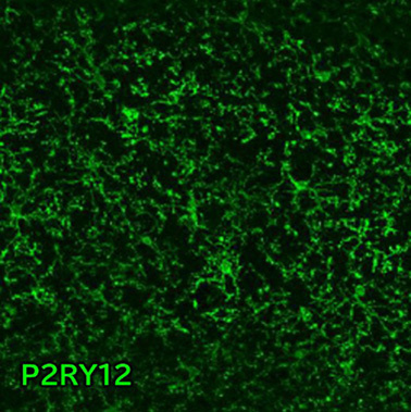
Immunohistochemistry was performed on mouse brain (frozen sections) using anti-P2RY12 and anti-Iba1 antibodies.
Data by courtesy of
Dr. Miyata, Department of Applied Biology, Kyoto Institute of Technology
Antibodies
P2RY12
Primary Antibody: Anti P2RY12, Guinea Pig (Product Number: 011-28873, This product) 1:1,000
Secondary Antibody: Alexa Fluor® 488 AffiniPure Goat Anti-Guinea Pig IgG (H+L) (Jackson ImmunoResearch, Product Number: 106-545-003)
Iba1
Primary Antibody: Anti Iba1, Rabbit (for Immunocytochemistry) (Product Number: 019-19741) 1:1,000
Secondary Antibody: Alexa Fluor® 594-AffiniPure Goat Anti-Rabbit IgG (H+L) (Jackson ImmunoResearch, Product Number: 111-585-003)
[Result]
The co-localization of P2RY12 and Iba1 signals was confirmed.
Differentiating between microglia and macrophages
In the cerebral cortex and the medulla oblongata area postrema, triple staining was performed using anti-P2RY12 antibody (microglia marker), anti-Iba1 antibody (microglia/macrophage marker), and anti-laminin antibody (basement membrane marker of cerebral blood vessels), and the localization of cells was confirmed.
Data by courtesy of
Dr. Miyata, Department of Applied Biology, Kyoto Institute of Technology
Antibodies
P2RY12
Primary Antibody: Anti P2RY12, Guinea Pig (Product Number: 011-28873, This product) 1:900
Secodary Antibody: Alexa Fluor® 488 AffiniPure Goat Anti-Guinea Pig IgG (H+L) (Jackson ImmunoResearch, Product Number: 106-545-003)
Iba1
Primary Antibody: Anti Iba1, Rabbit (for Immunocytochemistry) (Product Number: 019-19741) 1:500
Secondary Antibody: Alexa Fluor® 594-AffiniPure Goat Anti-Rabbit IgG (H+L) (Jackson ImmunoResearch, Product Number: 111-585-003)
Laminin
Primary Antibody: Anti Laminin Antibody (Rat, Made in Dr. Miyata’s Lab) 1:200
Secondary Antibody: DyLight™ 405-conjugated AffiniPure™ Goat Anti-Rat IgG (H+L) (Jackson ImmunoResearch, Product Number: 112-475-003)
[Result]
Among the Iba1-positive cells, those that are present in the brain parenchyma are P2RY12-positive and are thought to be microglia (arrowheads). On the other hand, Iba1-positive/P2RY12-negative cells are present around the blood vessel basement membrane, and it is thought that these cells are macrophages (arrows).
Comparison with anti-Iba1 antibody and anti-TMEM119 antibody
Immunohistochemistry was performed on mouse brain and spinal cord (frozen sections) using anti-P2RY12, anti-Iba1 and anti-TMEM119 antibodies
Antibodies
P2RY12
Primary Antibody: Anti P2RY12, Guinea Pig (Product Number: 011-28873, This product) 1:1,000
Secondary Antibody: Alexa Fluor® 647 AffiniPure Donkey Anti-Guinea Pig IgG (H+L) (Jackson ImmunoResearch, Product Number: 706-605-148)
Iba1
Primary Antibody: Anti Iba1, Goat (Product Number: 011-27991) 1:1,000
Secondary Antibody: Alexa Fluor® 488 AffiniPure Donkey Anti-Goat IgG (H+L) (Jackson ImmunoResearch, Product Number: 705-545-147)
TMEM119
Primary Antibody: Anti-TMEM119 Antibody (Rabbit, Competitor’s Product) 1:100
Secondary Antibody: Alexa Fluor® 594-AffiniPure Donkey Anti-Rabbit IgG (H+L) (Jackson ImmunoResearch, Product Number: 711-585-152)
[Result]
Compared to anti-TMEM119 antibody, our anti-P2RY12 antibody staining was able to detect fine protrusion structures more clearly.
Comparison with conventional product
Immunohistochemical staining was performed on mouse brain and spinal cord (frozen sections) using our company's and competitor’s anti-P2RY12 antibodies.
Antibodies
Fujifilm Wako
Primary Antibody: Anti P2RY12, Guinea Pig (Product Number: 011-28873, This product) 1:1,000 (Antibody concentration 0.5 μg/mL)
Secondary Antibody: Alexa Fluor® 488 AffiniPure Donkey Anti-Guinea Pig IgG (H+L) (Jackson ImmunoResearch, Product Number: 706-545-148)
Conventional product
Primary Antibody: Anti-P2RY12 Antibod (Rabbit, Competitor’s Product) 1:200 (Antibody concentration 0.5 μg/mL)
Secondary Antibody: Alexa Fluor® 488 AffiniPure Donkey Anti-Rabbit IgG (H+L) (Jackson ImmunoResearch, Product Number: 711-545-152)
[Result]
With the conventional product, nonspecific signals thought to be from neurons were observed (arrowheads). On the other hand, with this product, almost no nonspecific signals were observed.
Application Data
Immunohistochemistry
Mouse
Site: Hippocampus (CA2)
Sample: Frozen section
Antibody concentration: 1:200
Notes: Blue indicates DAPI.
Dr. I, F University
Site: Prefrontal cortex
Sample: Frozen section
Antibody concentration: 1:900
Notes: Blue indicates DAPI.
Dr. Yu, Graduate School of Medicine, Tohoku University
Site: Cerebral cortex
Sample: Frozen section
Antibody concentration: 1:10,000
Notes: Blue indicates DAPI.
Dr. Yamada, Faculty of Medical Sciences,
Kyushu University

Site: Hippocampus
Sample: Frozen section
Antibody concentration: 1:500
Dr. Koizumi and Dr. Saito,
Graduate School of Medicine, University of Yamanashi
/Yamanashi GLIA Center
Site: Hippocampus
Sample: Frozen section
Antibody concentration: 1:500
Dr. Hashimoto,
Institute for Human Life Science,
Ochanomizu University
Site: Hippocampus
Sample: Frozen section
Antibody concentration: 1:1,000
Notes: Activated treatment (sodium citrate buffer)
Dr. Hashimoto,
Institute for Human Life Science,
Ochanomizu University
Site: Hippocampus
Sample: Paraffin section
Antibody concentration: 1:1,000
Notes: Activated treatment (sodium citrate buffer, pH 6.0, 80 °C, 15 minutes)
Dr. Sahara,
Brain Research Institute Center for Integrated Human Brain Science, Department of Functional Neurology and Neurosurgery, Niigata University
Site: Cerebral cortex
Sample: Paraffin section
Antibody concentration: 1:1,000
Notes: Activated treatment (sodium citrate buffer, pH 6.0, 80 °C, 15 minutes)
Dr. Sahara,
Brain Research Institute Center for Integrated Human Brain Science, Department of Functional Neurology and Neurosurgery, Niigata University
Site: Median eminence
Sample: Frozen section
Antibody concentration: 1:1,000
Dr. Furube,
Department of Functional Anatomy and Neuroscience,
Asahikawa Medical University
Site: Spinal cord
Sample: Frozen section
Antibody concentration: 1:2,000
Dr. Tsuda and Dr. Kohno,
Department of Molecular and System Pharmacology,
Graduate School of Pharmaceutical Sciences,
Kyushu University

Site: Trigeminal ganglion
Sample: Frozen section
Antibody concentration: 1:900
Note: S-100β is a marker for satellite glial cell (SBC)
Site: Retina
Sample: Paraffin section
Antibody concentration: 1:400
Notes: Activated treatment (TE buffer, pH9.0, 3 minutes of pressure)
Dr. Ishizuka,
Department of Pathophysiology and Metabolism,
Kawasaki Medical School
Rat
Site: Cerebral cortex
Sample: Frozen section
Antibody concentration: 1:500
Notes: Activated treatment (pH6.0 citrate buffer, 75℃, 20 minutes)
Blue indicates DAPI.
Dr. Koizumi, Anatomy and Neurobiology,
Kyoto Prefectural University of Medicine
Please note that Fujifilm Wako cannot answer any questions regarding the data above.
References
- Butovsky, O. et al.: Neurosci., 17(1), 131(2014).
Identification of a unique TGF-β–dependent molecular and functional signature in microglia - Keren-Shaul, H. et al.: Cell, 169(7), 1276(2017).
A unique microglia type associated with restricting development of Alzheimer’s disease - Maeda, J. et al.: Brain Commun., 3(1), fcab011(2021).
Distinct microglial response against Alzheimer's amyloid and tau pathologies characterized by P2Y12 receptor
FAQ
About antibody
- What is the antigen?
- It is a synthetic peptide of P2RY12 (corresponding to the C-terminus). The specific sequence is not disclosed.
About protocol
- What secondary antibodies can be used?
- At Fujifilm Wako, the following secondary antibodies have been successfully used:
Alexa Fluor® 488 AffiniPure Goat Anti-Guinea Pig IgG (H+L) (Jackson ImmunoResearch, Product Number: 106-545-003)
Alexa Fluor® 488 AffiniPure Donkey Anti-Guinea Pig IgG (H+L) (Jackson ImmunoResearch, Product Number: 706-545-148)
Alexa Fluor® 647 AffiniPure Donkey Anti-Guinea Pig IgG (H+L) (Jackson ImmunoResearch, Product Number: 706-605-148)
About application
- What animals can this antibody be used for?
- Fujifilm Wako has confirmed that it can be used for mice. There are also reports of staining in rats, although we have no experience of this at our company. For details, please refer to the “Data” section. Other species have not been tested.
- What kind of tissues or sites can be stained?
- Fujifilm Wako have experience in staining the following tissue and site:
- Cerebral cortex
- Hippocampus
- Hypothalamus
- Cerebellum
- Olfactory bulb
- Spinal cord
- Prefrontal cortex
- Medulla oblongata area postrema
- Median eminence
- Trigeminal ganglion
- Retina
- Can paraffin sections be stained?
- Fujifilm Wako has not confirmed the application of immunohistochemical staining of paraffin sections using this antibody. However, the staining results have been reported in paraffin sections of mouse hippocampus and cerebral cortex. For details, please refer to the“Data” on this page.
- Can microglia other than homeostatic microglia be stained?
- P2RY12 is a marker for homeostatic microglia, and it is known that the expression level of P2RY12 is reduced in disease-associated microglia (DAM) and microglia in Alzheimer's disease model mice2-3). In these microglia, It is possible that these microglia cannot be stained due to the reduced expression.
- Can satellite glial cells be stained?
- There is the report of co-staining with S-100β in the trigeminal ganglion of mice. For details, please refer to the“Data” on this page.
About troubleshooting
- The background of immunohistochemistry is high.
- Try glycine treatment (In 25 mM glycine/PBS for about 20 minutes) after preparation of frozen sections. It is possible to inactivate the aldehyde groups in the sample and suppress the background.
Overview / Applications
| Outline | P2RY12 (also known as P2Y12 or P2Y12R) is an adenosine 5'-diphosphate (ADP) receptor and a member of the P2Y family of purinergic G protein-coupled receptors. In the central nervous system, it is specifically expressed in microglia and is used as a microglial marker. Anti P2RY12, Guinea Pig is a guinea pig polyclonal antibody that reacts with P2RY12. [Antigen] Synthetic peptide corresponding to the C-terminus of P2RY12 [Species cross reactivity] Mouse(Other species have not been tested) [Application] Immunohistochemistry (frozen section) 1:500-1:2,000 |
|---|---|
| Precautions for Use | Avoid repeated freeze and thaw. |
Property
| Appearance | Liquid |
|---|---|
| Concentration | Protein : 0.45 - 0.65 mg/mL |
Manufacturer Information
Alias
- Anti P2Y12, Guinea Pig
For research use or further manufacturing use only. Not for use in diagnostic procedures.
Product content may differ from the actual image due to minor specification changes etc.
If the revision of product standards and packaging standards has been made, there is a case where the actual product specifications and images are different.
The prices are list prices in Japan.Please contact your local distributor for your retail price in your region.


