Nucleic Acid Staining Reagents
A nucleic acid staining reagent is used for visualization of nucleic acids in a gel and for analysis of nucleic acids separated by electrophoresis. Ethidium bromide is widely used staining reagent in nucleic acid staining and is positioned as a standard for this purpose. However, as ethidium bromide has a mutagenic potential, caution should be exercised when handling it. Therefore, a new, highly safe nucleic acid staining reagent, SAFELOOK™, has recently been developed. Fujifilm Wako provides both ethidium bromide and highly safe nucleic acid reagents.
Product Line-up
More Information
Principles of Nucleic Acid Staining Reagents
A compound used as a nucleic acid staining reagent has the property that its fluorescence intensity increases when it is bound to nucleic acid. Commonly used ethidium bromide (EtBr) intercalates into double-stranded DNA, which increases the fluorescence intensity by about 20 times (it also binds to single-stranded nucleic acids).
Widely used nucleic acid staining reagents other than ethidium bromide include 4',6-diamidino-2-phenylindole (DAPI), propidium iodide (PI), and acridine orange (AO).
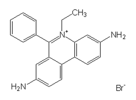
Mutagenicity of ethidium bromide
Nucleic acid staining reagents may induce mutations due to their property of binding to nucleic acids. Actually, ethidium bromide has been determined to be mutagenic by the Ames test1), and adequate caution is needed when handling it. Fujifilm Wako provides highly safe nucleic acid reagents SAFELOOK™.
Selection of Staining Method
Nucleic acid staining methods for agarose electrophoresis are classified into prestaining, post-staining, and add-to-sample methods. The strengths and weaknesses of each method are summarized below.
| Staining method | Strengths | Weaknesses |
|---|---|---|
| Prestaining A staining reagent is added to agarose gel in advance. Suitable when the results are needed quickly and conveniently |
•Time for staining is not required after electrophoresis. •Bands can be observed during electrophoresis. |
•When there is a difference in DNA concentration, the positions of bands of the same molecular weight may differ among lanes, due to variation in binding of staining reagent. •After prolonged electrophoresis, the staining reagents can run off the gel (when EtBr is used) |
| Post-staining After electrophoresis, the agarose gel is soaked in buffer containing the staining reagent Suitable when accuracy of the results is most important |
•Variation in DNA concentration less likely disrupt the electrophoresis pattern. •Electrophoresis can be performed for a long time. •Gels containing denaturant can be stained. (buffer replacement is required) |
•Time for staining is required after electrophoresis. •Bands cannot be observed during electrophoresis. |
| Add-to-sample A staining reagent is added to a sample to be analyzed by electrophoresis Suitable when the results are needed quickly and conveniently |
•Time for staining is not required after electrophoresis. •Bands can be observed during electrophoresis. |
•When there is a difference in DNA concentration, the positions of bands of the same molecular weight may differ among lanes, due to variation in binding of staining reagent. •After prolonged electrophoresis, the staining reagents can run off the gel. |
References
- Huang, Q., & Fu, W. L.: Clin. Chem. Lab. Med., 43(8), 841(2005).
Comparative analysis of the DNA staining efficiencies of different fluorescent dyes in preparative agarose gel electrophoresis
For research use or further manufacturing use only. Not for use in diagnostic procedures.
Product content may differ from the actual image due to minor specification changes etc.
If the revision of product standards and packaging standards has been made, there is a case where the actual product specifications and images are different.
The prices are list prices in Japan.Please contact your local distributor for your retail price in your region.